A new paper, written by a team including Iconeus staff and published in the journal eBiomedicine, shows for the first time that our 3D ultrasound localization microscopy (ULM) technology can be used to image the whole-brain microvasculature in the mouse, while keeping the skull intact. Using this recently-developed functional ultrasound approach, we were able to achieve a resolution down to 20 µm – an unprecedented achievement for an in vivo technique.
Being able to image blood vessels and blood flows in the brain at high resolution is essential for studying a wide range of brain disorders, including aneurysm, stroke and degenerative diseases. However, current imaging modalities either have limited spatial resolution or poor penetration depth. We’re therefore excited to report the publication of a new paper that pushes the boundaries of what is possible for in vivo mouse brain imaging, using our 3D ultrasound localization microscopy (ULM) technology.
Ultrasound localization microscopy
ULM works by using fUS to detect and track injected microbubbles as they travel through the brain’s blood vessels. The movements of the bubbles allow us to build up fantastically detailed images of the brain microvasculature, with resolution as low as 5 µm.
For over 5 years now, we’ve been using this method to generate 2D images of the brain, but of course blood vessels are oriented in three dimensions. Although several approaches have been proposed, up to now none of them has been used to image the mouse brain through the intact skull, but we’ve been working on a 3D ULM method that can do just that.
High-resolution 3D imaging in vivo
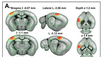
The new paper, published in the journal eBiomedicine (part of The Lancet Discovery Science), describes how we used our 3D ULM approach to track 3 million microbubbles with a temporal resolution of 750 volumes per second. This allowed us to determine flow velocities in the range 2–100 mm/s, meaning that we were able to visualize everything from large vessels (∼100 µm diameter) to capillaries (down to 20 µm diameter). The smaller vessels imaged are very important in regulating blood flows and brain perfusion, and so the technique has immediate application interest.
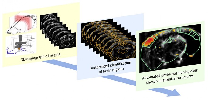
As in our previous work, the blood vessels imaged using this 3D ULM method were automatically registered to the Allen Mouse Brain Atlas. Using our IcoStudio software, this meant that we could extract microbubble tracks for quantification from any region of the brain that we wanted to.
Potential neuroscience applications
Ludovic Lecointre, Pharm.D., CEO and co-founder of Iconeus, emphasized the significance of what has been achieved by the Iconeus team: “Imaging the vascular networks in the whole brain remains really hard to do in vivo, because it spans such a wide range of scales and velocities. For example, MR and CT-based micro-angiography using contrast agents are limited to showing the anatomy, and don’t provide the time dynamics needed for assessing cerebrovascular function. On the other hand, optical imaging provides blood flow information with microscale-level resolution, but can’t show structures below the surface layer of the brain”.
“By carrying out this study, we’ve shown the great potential of the 3D ULM approach for generating images of the whole-brain vasculature at unprecedented scale for an in vivo technique. As a result, we think it should facilitate brain research in many areas – from vascular disease progression and vascular remodeling to tumor growth and efficacy of therapeutic interventions. I’m really looking forward to future developments and applications of this technique”.
Reference:
O. Demeulenaere, A. Bertolo, S. Pezet, N. Ialy-Radio, B. Osmanski, C. Papadacci, M. Tanter, T. Deffieux and M. Pernot, In vivo whole brain microvascular imaging in mice using transcranial 3D Ultrasound Localization Microscopy, eBiomedicine, 2022, 79: 103995.