A collaboration between Paris-based academic researchers and major US pharma company Biogen has used fUS for the first time to investigate functional responses in a mouse model of multiple sclerosis. They found, as expected, that regions with higher levels of demyelination showed higher blood flows following whisker stimulation, laying the groundwork for more in-depth studies on the link between neurovascular health and Multiple Sclerosis.
Multiple sclerosis (MS) is an autoimmune disease of the central nervous system that affects about 2.8 million people worldwide. One feature of MS is demyelination – the deterioration of the fatty myelin sheaths surrounding the nerve fibers (axons) of neurons, which causes a reduction in the speed and efficiency of signal transmission.
Although treatments are available to manage the symptoms of MS, there is currently no cure, and considerable effort has been invested on this front. A key obstacle in this effort has been the absence of reliable early biomarkers – especially in preclinical research – which are essential for detecting the disease at its earliest stages and for evaluating the efficacy of potential treatments. Up to now, researchers have used variants of MRI, PET or electrophysiology, which although valuable in many ways, fail to simultaneously address issues of resolution, depth of field and practicality in small animals, especially when wishing to dynamically assess brain function as opposed to obtaining static images of the vasculature.
Assessing neurovascular blood flow
A way of overcoming the above-mentioned limitations has now been outlined in a new study carried out by a team led by Sophie Pezet at Physics for Medicine Paris, along with collaborators at major US pharma company Biogen and Sorbonne University – including Iconeus co-founders Mickael Tanter and Thomas Deffieux.
The work, reported in the journal Imaging Neuroscience, describes the use of functional ultrasound (fUS) to study a mouse model of MS. Four groups of eight adult mice were studied over a period of eight weeks, with three of the groups given cuprizone to induce demyelination, and with the other group used as a control.
Using an Iconeus One system fitted with an IcoPrime probe, the anesthetized mice were imaged during a period of calibrated whisker stimulation. In this way, the extent of increased brain blood flow could be assessed, with the images automatically registered to the Allen Mouse Brain Atlas using IcoStudio software. At the end of the study, the mice were euthanized, and brain slices stained to assess levels of myelin basic protein (MBP), a key marker for intact myelin.
Reliable metrics, surprising responses
On the basis of the fUS data, the researchers uncovered clear changes in brain function in the cortex, (as assessed by an increase in the number of active pixels), a gradual rise in steady-state cerebral blood volume within these pixels, and an increase in ‘rise time’ (a measure of the speed of response). These three metrics were also well-correlated with the level of MBP, confirming that fUS can be used to give a reliable measure of demyelination.
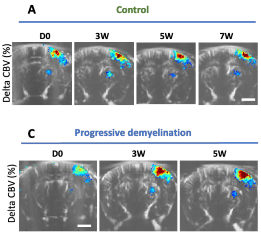
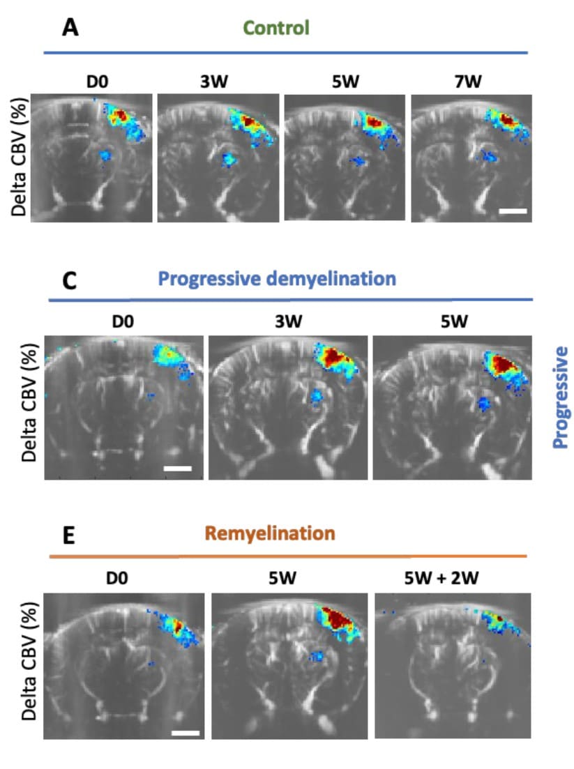
Example fUS images from the mouse study, over a period of 0–7 weeks, showing the change in cerebral blood volume (CBV) following whisker stimulation in (A) a control animal and (C) an animal treated with cuprizone and exhibiting the demyelination characteristic of MS; note the increased response at 3 weeks and 5 weeks. Reproduced from Imaging Neuroscience and published under CC BY 4.0.
The increased responses seen in this work are mirrored in previous clinical studies, and the authors say that several researchers “have hypothesized that these surprising, enhanced responses might be an adaptive plasticity to compensate for the growing disability of the diseased brain”. They also point out that it remains unclear whether these vascular events are indeed a consequence of a disease onset, or whether they may actually be the primary cause.
A further finding was that introducing small demyelinating lesions into specific regions did not result in a change in hemodynamic response, leading them to conclude that local neuroinflammation and demyelination on their own are insufficient to induce these response changes.
Pushing the boundaries of preclinical research
The results raise numerous questions for future study, and the authors point out the desirability of carrying out studies over longer periods of time, in order to determine whether responses ultimately stop increasing and start to decrease, as has been found in MS patients. As the authors have found in this work, uncovering insights is made considerably easier by the ease with which longitudinal studies on single animals can be carried out using fUS, in contrast to other techniques.
In their concluding remarks, the authors mention the “high sensitivity” achievable with fUS-based measurements of blood volumes, and say that their study “underscores the potential of using altered hemodynamic responses as a correlate for myelin loss in this model”.
Bruno Osmanski, Ph.D., Co-founder & Chief Scientific Officer at Iconeus, says that “From our perspective at Iconeus, this study is certainly an interesting and valuable application of fUS, showing – once again – that the technique continues to be used to push the boundaries of knowledge in all areas of preclinical research”.
The involvement of the major US pharma company Biogen in this work has been especially welcome, because it highlights how fUS is being taken increasingly seriously across the sector. With further collaborations between academia and industry like this, researchers should be in a good position to make progress on understanding the link between neurovascular health and MS – which as a serious disease affecting so many people is very much in need of more effective treatments”.
Reference:
B. Beliard, L. Delay, Y. Travert-Jouanneu, N. Ialy-Radio, C. Isaad, A. Réaux-Le Goazigo, T. Deffieux, D.P. Bradley, M. Tanter and S. Pezet, Novel insights into vascular dysfunction in cuprizone-induced demyelination through functional ultrasound imaging, Imaging Neuroscience, 2025, 3: article no. 00534, DOI: https://doi.org/10.1162/imag_a_00534.