Clinical research
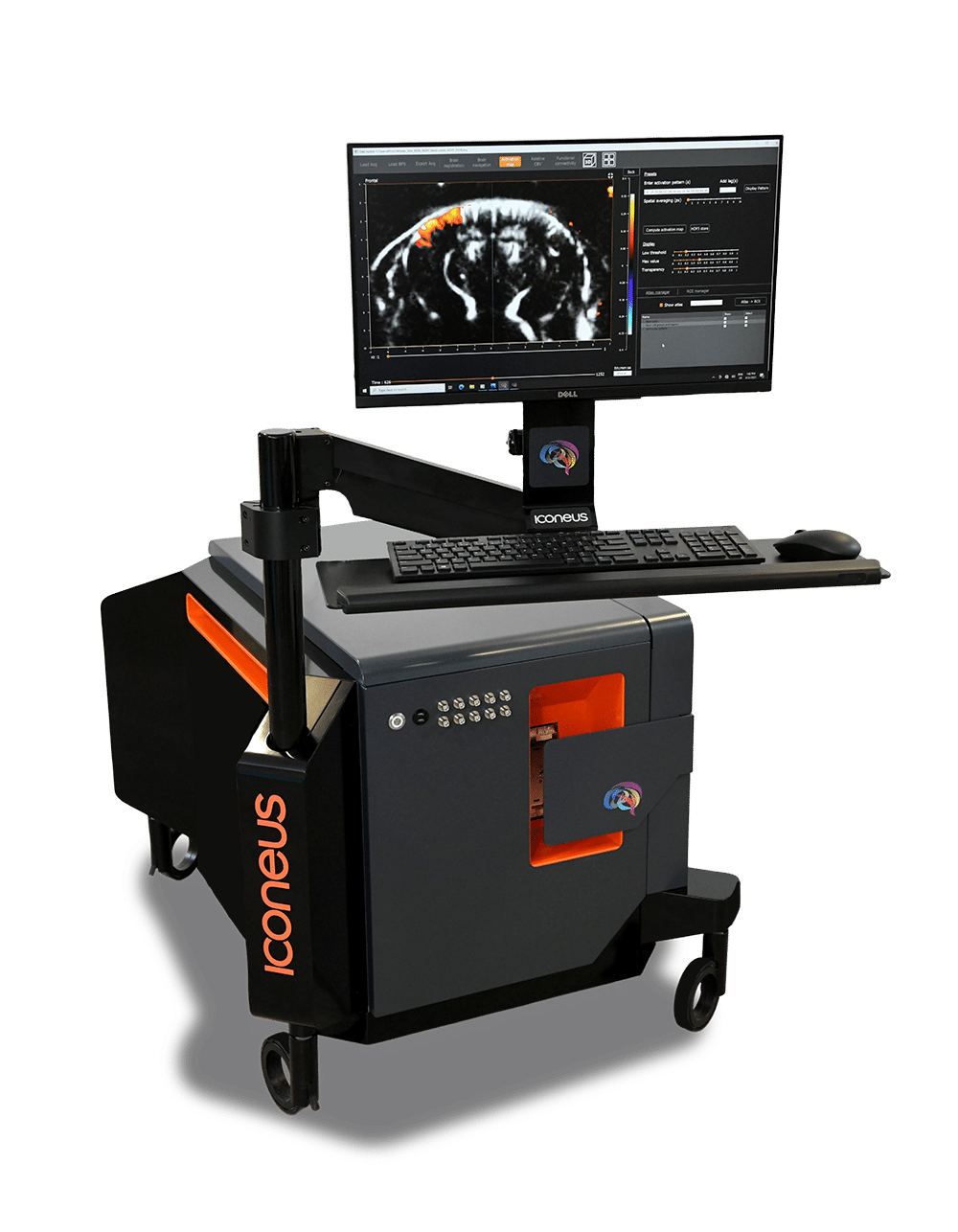
Building on years of preclinical expertise and research, we’re now investing in and supporting exciting new clinical applications, using our Iconeus One Plus CR functional ultrasound system dedicated to clinical research.

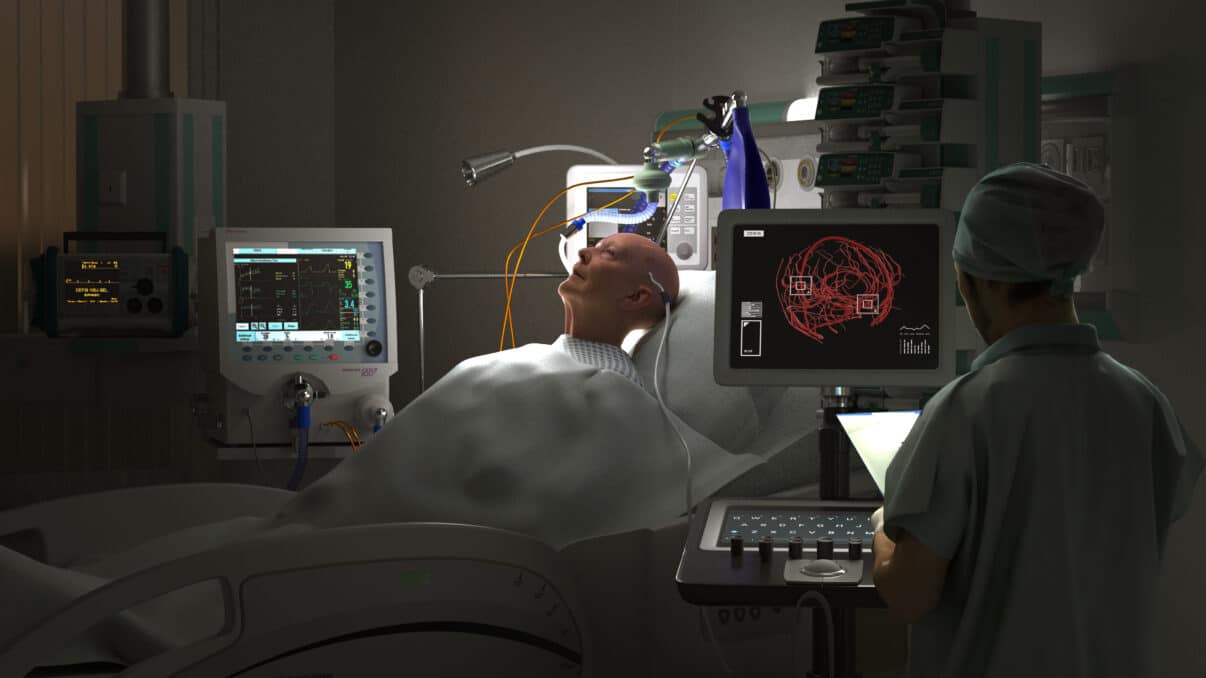
Improving diagnosis of stroke and aneurysm
Detecting ischemia for improved treatment of traumatic brain injury and stroke
Developing whole-brain transcranial functional ultrafast localization microscopy in adults
A key challenge in dealing with strokes is distinguishing between ischemic and hemorrhagic stroke as early as possible, as well as monitoring aneurysm. A promising approach to achieve this is transcranial imaging of deep vasculature (and quantification of hemodynamic parameters).
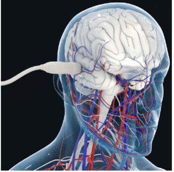
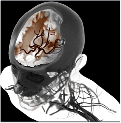
This is now possible in the adult human brain at microscopic resolution using ultrasound localization microscopy (ULM) of intravenously injected microbubbles. To take advantage of this concept, we’re developing a transcranial ULM scanner for stroke and aneurysm diagnosis in adults, in close collaboration with Geneva University and Physics for Medicine Paris.
Brain damage resulting from traumatic brain injury or subarachnoid hemorrhage is a major cause of death and severe disability, and secondary ischemic damage is a significant contributor to morbidity. Optimizing cerebral blood flows can help prevent this secondary damage, but detecting the signs at a sufficiently early stage remains challenging.
In conjunction with Caen University Hospital, the Physiopathology and Imaging of Neurological Disorders (PhIND) group based in Caen, and Physics for Medicine Paris, we’re developing a small implantable probe using our ultrafast Doppler technology, with the aim of allowing continuous cerebral blood flow monitoring in several brain regions for brain-injured patients.
This is a ‘moonshot’ project that is focused on developing a highly temporally-resolved version of our existing ultrafast localization microscopy (ULM) technology for Iconeus One. In this clinical proof-of-concept project, we are initially aiming to achieve diagnosis and treatment monitoring of cerebral small vessel disease, stroke, aneurysms, and alterations of the neurovascular response.
To reduce the temporal resolution from the current 60 s to the <1 s needed for this ‘functional ULM’ (fULM) technology, we’re planning to use a set of highly sensitive ultrasound probes within a helmet. This will present significant challenges, but in conjunction with our collaborators in the EU-funded MICROVASC project, we’re confident about making progress, and are looking forward to seeing what can be achieved for clinical applications.
Assessing neurodevelopmental disorders in newborns
Neurodevelopmental disorders are a major public health issue, but there is currently no effective imaging modality to assess the brain function of at-risk newborns: functional MRI is expensive and difficult to access, and EEG and NIRS only provide surface images. As a result, diagnosis is often made once cognitive and behavioral changes are apparent (at the age of two or three), which limits therapeutic approaches.
Working for the CONEXUS project (a collaboration between the Robert Debré Hospital and Physics for Medicine Paris), we’re committed to developing a brain imager for newborns based on functional ultrasound imaging. The goal will be to assess brain functional connectivity at a baby’s bedside over a period of time, ultimately transforming neonatology services and providing early biomarkers of neurodevelopmental disorders.
To learn more about this project, read our news release.
Disclaimer
The ultrasound devices presented are not CE‑marked and are not FDA‑cleared, and must not be used for any clinical purpose.
Any investigational use requires prior authorization from the competent regulatory authority and supervision by qualified professionals.
All information is provided for informational purposes only and does not constitute medical advice.
Iconeus On
The only fUS system designed specifically for neuroscientists
Iconeus One is an imaging system with a difference – one with the sensitivity and resolution needed to see what’s happening in the brain at the finest scales. Not only does it work on awake or moving animals, but you can even see what’s happening in real-time.
Contact us
Interested by what you’ve read about functional activation mapping using fUS? Talk to one of our specialists about your application.
Other applications