Resting-state functional connectivity
In an analogous fashion to fMRI, functional ultrasound can be used to map intrinsic brain connectivity (often called resting-state functional connectivity or resting-state networks), by detecting correlated fluctuations of spontaneous blood flow.
The high spatial and temporal resolution of fUS, and its much greater inherent sensitivity than BOLD-based fMRI, makes it a powerful way of studying neuropsychiatric diseases, and potentially aiding early diagnosis. Below are a few examples of fUS in action.

2D connectivity matrices
3D connectivity matrices
Seed-based correlation mapping
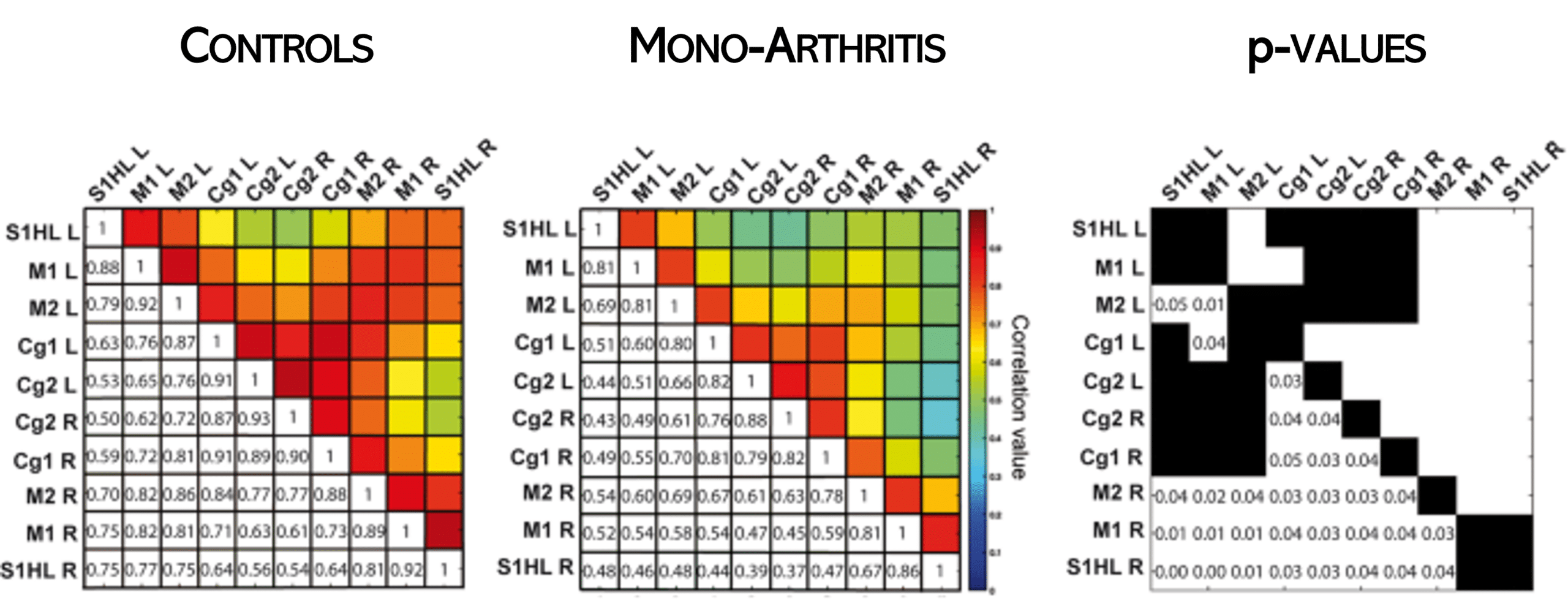
Functional connectivity in pathological models
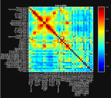
Generating connectivity matrices in two dimensions involves investigating responses from a single coronal plane, as illustrated here for a mouse model.
This task is difficult using fMRI, because the small size of the mouse brain demands expensive, high-field fMRI scanners, and because maintaining stable physiological parameters (and hence efficient neurovascular coupling) is very difficult in anesthetized mice. In addition, the reliability of a connectivity matrix depends critically on accurately defining the ‘region of interest’ (ROI).
Doing this using Iconeus One is quick and reliable, because it allows automated correlation to the Allen Mouse Brain Atlas, meaning results are much less vulnerable to bias.
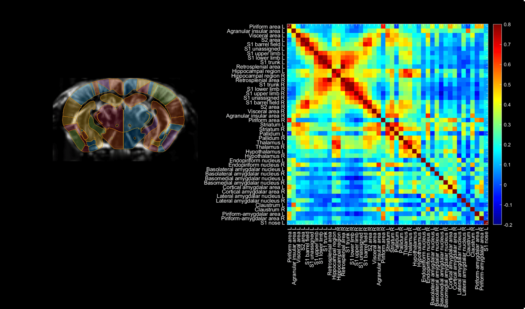
3D connectivity matrices are simply an extension of the 2D approach mentioned above – in essence, you’re determining the overall connectivity between (usually) closely-spaced slices.
The fast switching between planes used in Iconeus One shortens the whole process to 20 minutes or less.
Another way of visualizing brain activity relationships is by correlating voxel activity across the whole brain with the activity of a small ‘seed’ region – known as seed-based (or ROI-based) functional connectivity.
This approach is not so dependent on the segmentation of the brain into functional regions, since you only need to define a single region.
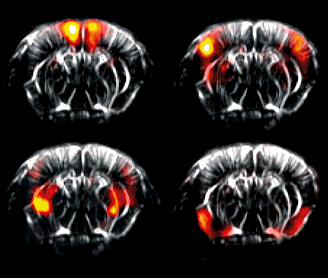
A different manifestation of functional connectivity is looking at how differences relevant to animal pathologies influence connectivity across the whole brain.
For example, functional ultrasound has been used to study the role of oxytocin in rat pups (Mairesse et al., Glia, 2019) and to investigate sensitivity to inflammatory pain in anesthetized rats, as shown here.
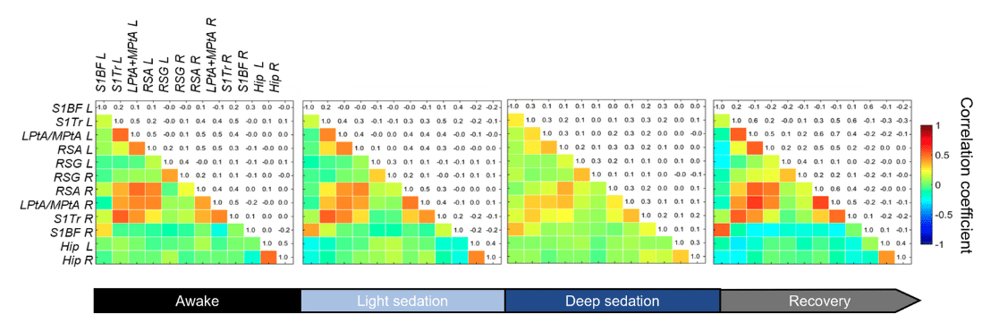
Investigating connectivity in moving animals
Connectivity matrices are normally developed for anesthetized animals, but an important development is using the light, robust Iconeus One probes to obtain results from awake mice (freely-moving or head-fixed), eliminating the bias of anesthetics.
As shown in the example below, this capability allows the effects on brain activity of anesthetics themselves to be studied, and it may also be relevant for analyzing genetically modified mice models.
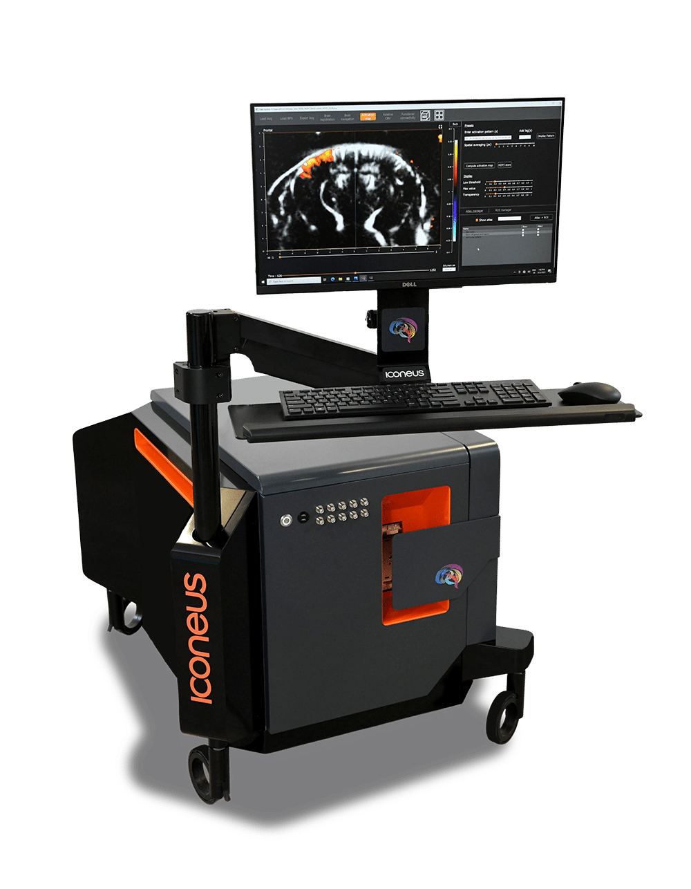
Iconeus One
The only fUS system designed specifically for neuroscientists
Iconeus One is an imaging system with a difference – one with the sensitivity and resolution needed to see what’s happening in the brain at the finest scales. Not only does it work on awake or moving animals, but you can even see what’s happening in real-time.
Contact us
Interested by what you’ve read about functional activation mapping using fUS? Talk to one of our specialists about your application.
Other applications