First study published on clinical use of Iconeus fUS equipment
A major milestone for Iconeus: The first clinical study using Iconeus One
In a major milestone for neurological research and for Iconeus, we report on the use of our equipment for fUS imaging of the spinal cord in human subjects.
A new paper published in the journal Neuron reports on the use of fUS to image the spinal cord in human subjects. This is a major milestone not just because it aids understanding of a key treatment option for chronic back pain, but because it is the first published study on the use of our fUS equipment in a clinical setting.
Injury or disease affecting the spinal cord can lead to interruption of electrical signals, resulting in a range of negative impacts on health, including chronic back pain. A variety of techniques are used to address these problems, amongst which an electrotherapy approach known as epidural spinal cord stimulation (ESCS) has shown particular promise.
However, in common with other treatments, ESCS doesn’t work for all patients, meaning that researchers are motivated to understand its mode of action and to predict which patients are likely to benefit the most.
Unfortunately, the spinal cord is a challenging target for imaging technologies, not least because of its small diameter of just 10–15 mm. Moreover, imaging the thoracic and lumbar segments of the spinal cord is particularly difficult using conventional techniques such as fMRI, because motion artefacts can arise by proximity to the lungs, and nearby tissue interfaces can cause magnetic effects. All these factors have hampered an understanding of its functional anatomy, limiting the effective use of treatments such as ESCS.
Detecting hemodynamic activity in the spinal cord
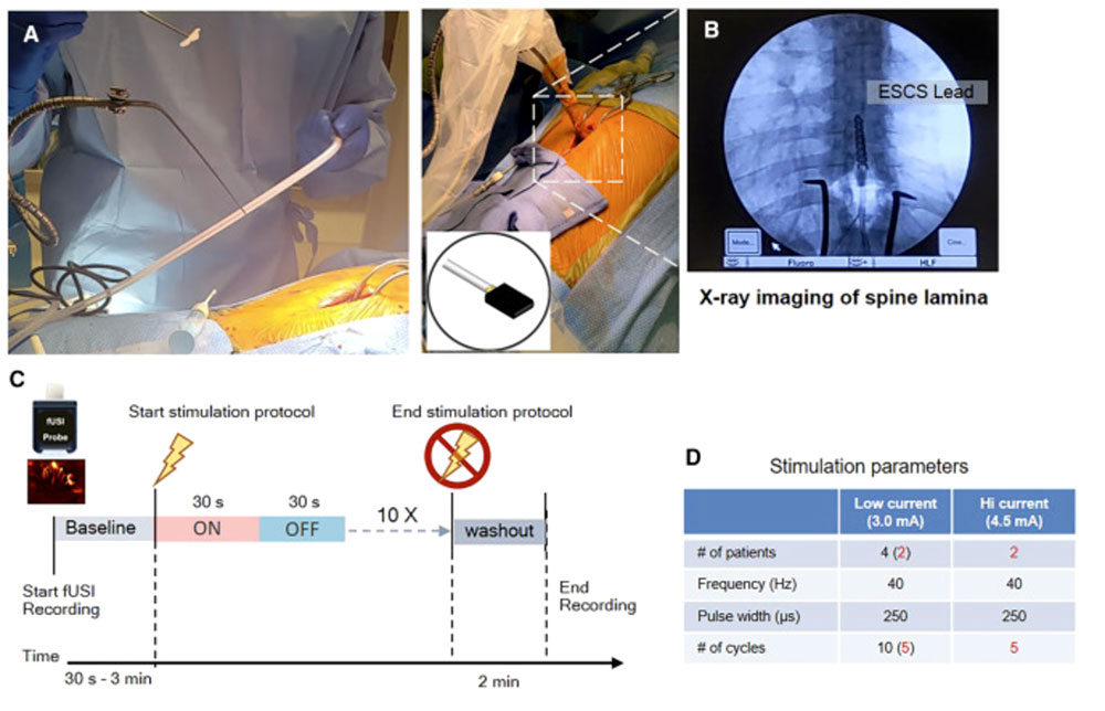
But all this could be about to change, as a result of work jointly led by Vassilios N. Christopoulos at the University of California Riverside, together with V. Reggie Edgerton and Charles Liu at the Keck School of Medicine, Los Angeles. In their work, now reported in the journal Neuron, they describe the use of functional ultrasound (fUS) to probe, for the first time, the functional changes of the human spinal cord in response to ESCS.
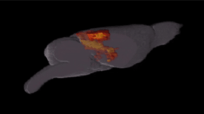
Using an Iconeus One system, the authors monitored the hemodynamic activity of the 10th thoracic vertebra through laminar openings of the spinal cord in four patients who had been implanted with an ESCS device to treat chronic back pain. The resulting strong hemodynamic activity detected, they say “provides the first evidence that [fUS imaging] can capture and characterize evoked changes in spinal cord blood volume dynamics in humans”.
The authors went on to use machine learning techniques to extract signals most strongly associated with stimulation, enabling them to predict the effects of ESCS on the hemodynamics within the spinal cord. They conclude that “Overall, our findings open new avenues to understand spinal cord function” and say that it has “considerable potential” for gaining a better understanding of the mechanism of action of ESCS.
The benefits of fUS for studying spinal cord function
From our perspective, the study is not only valuable for the reasons mentioned above, but because of its significance as the first publication relating to a clinical study using Iconeus One.
In addition, the authors point out several features of fUS that offered advantages in their work. One of these was the ability to detect regional hemodynamic changes of only 2% without averaging over multiple trials, which they say is necessary for using it to monitor moment-to-moment hemodynamic changes.
They also say that “its superior spatiotemporal performance and sensitivity offer substantially closer connection to the underlying neuronal signal than achievable with other hemodynamic methods such as fMRI”. This feature of fUS of course offers advantages across a range of neuroscientific fields, including but certainly not limited to spinal cord investigations.

Ludovic Lecointre, Pharm.D., Co-founder & Chief Executive Officer of Iconeus, said: “As the authors say themselves, this represents, a ‘major leap’ in the use of fUS, and we’re delighted to have supplied the technology that made it possible. Spinal cord neuroimaging has long been a very difficult technical challenge, making these results all the more significant. It therefore sets the stage for future clinical research investigations in this important area, as well as being a milestone in the use of fUS!”.
Reference:
K.A. Agyeman, D.J. Lee, J. Russin, E.I. Kreydin, W. Choi, A. Abedi, V.R. Edgerton, C. Liu and V.N. Christopoulos, Functional ultrasound imaging of the human spinal cord, Neuron, 2024, https://doi.org/10.1016/j.neuron.2024.02.012