
Iconeus One
The only functional ultrasound system designed specifically for neuroscientists
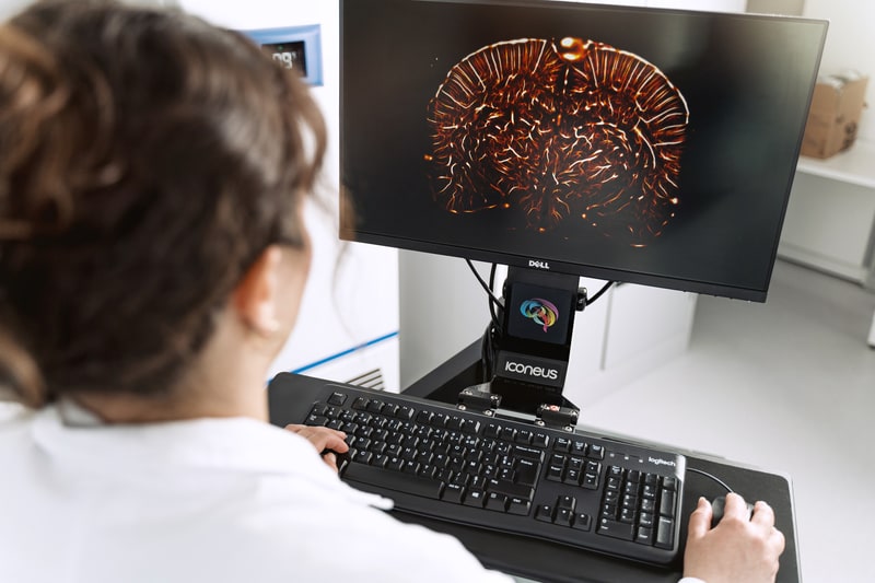
Iconeus One is an imaging system with a difference – one with the sensitivity and resolution needed to see what’s happening in the brain at the finest scales. Not only does it work on awake or moving animals, but you can even see what’s happening in real-time.
And we’re well aware that you don’t want to spend ages learning how to use it. So we’ve taken special efforts to make the software and hardware easy to use – and of course customizable to your specific needs.
 Powered by Bioz
What you get with Iconeus One
Here’s what’s available with our standard Iconeus One package
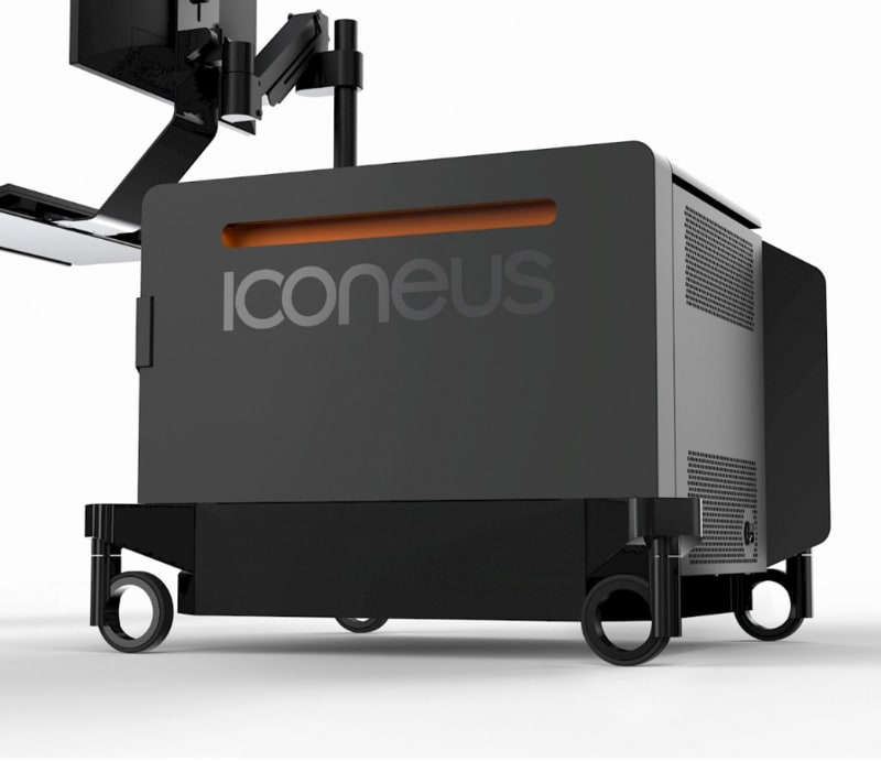

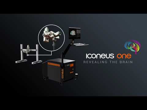
A portable imaging station, featuring our plane-wave ultrasound technology.
A height-adjustable keyboard and monitor.
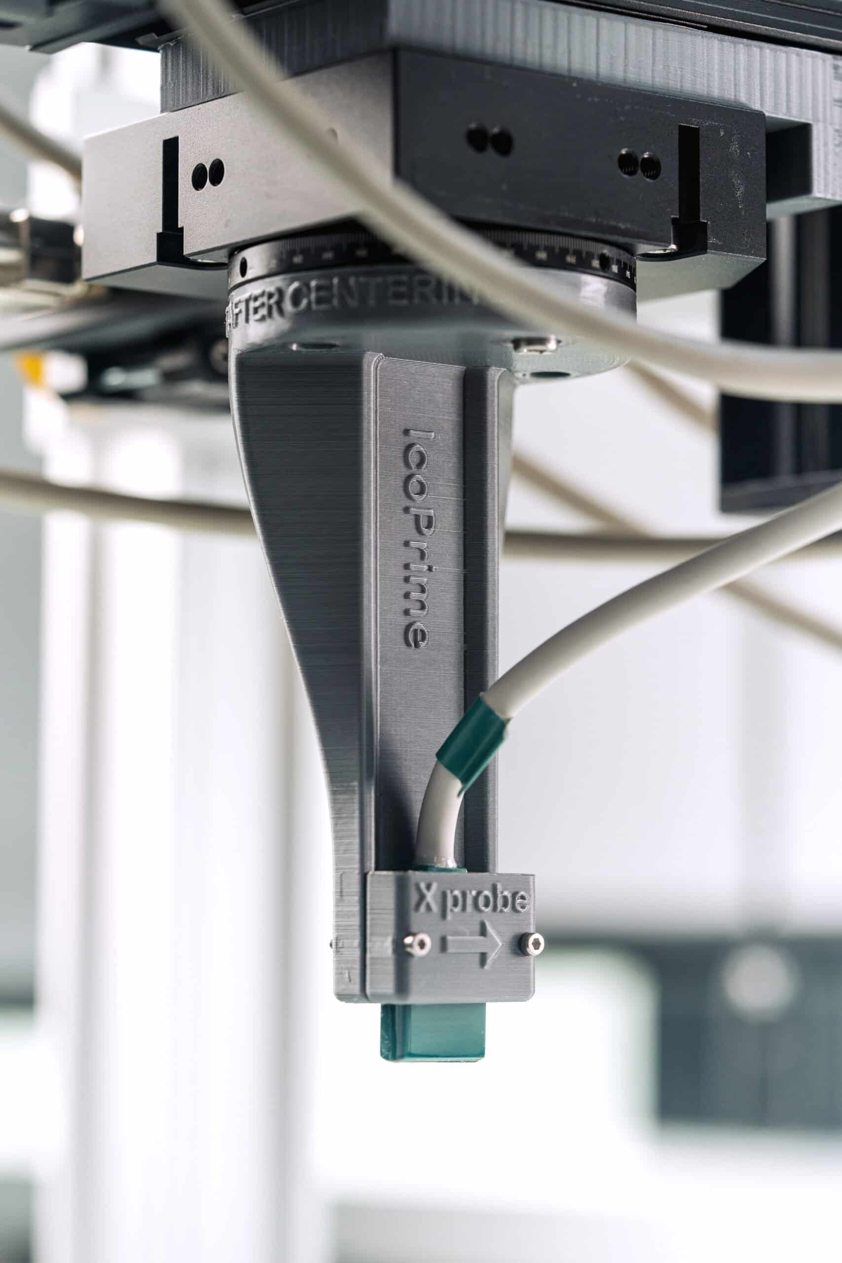
An ultrasound probe, optimized for the animal model you're studying (in a mobile or head-fixed configuration).
A four-axis motorized scanning platform for imaging head-fixed animals.
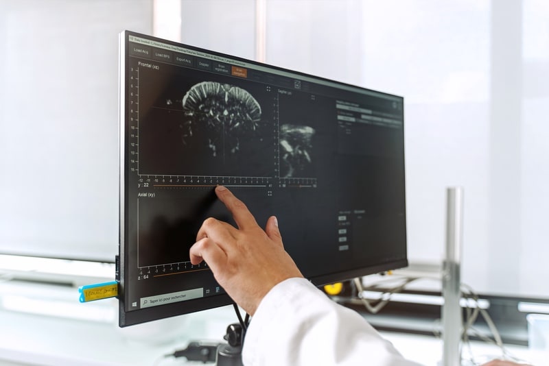
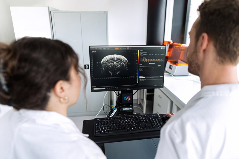
Easy-to-use software with an interactive live viewer, integrated adult mouse brain
Reference atlas, and three additional licenses for off-line data analysis.
Installation, training, ongoing support, and a one-year extended warranty.

Discover our Iconeus One brochure
Probes, robotic automation, software and more…
Our plane-wave functional ultrasound technology lies at the heart of the success of Iconeus One.
But the performance of the whole system depends on some other innovations too, including:
- Low-noise, ultra-light probes that are easily interfaced with the animal
- A robotic platform with four dimensions of movement (X, Y, Z and rotation) for in-depth, automated scanning
- Highly parallel ultrasound electronics, allowing simultaneous acquisition of large amounts of data and efficient removal of non-vascular signals from surrounding tissues
- Easy-to-use software, with a live viewer for instant insights and a reference atlas for the adult mouse brain.
Main features
Awake imaging
3D scans
High sensitivity
Super resolution
Plug and play
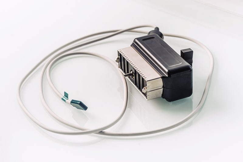
fUS probes optimized for your application
What you get from functional ultrasound depends crucially on the specifications of the probe: regular ultrasound transducers simply aren’t good enough for brain imaging.
So we’ve spent a lot of time maximizing the sensitivity of our probes, minimizing their size for mobile configurations, and working out the best specifications – including optimal frequencies – for different animal models.
Ultimately, we know that as a researcher, you want to keep ahead of your field – and for that you need a functional ultrasound system that’s optimized for your animal and your application. So we’ll be delighted to hear about your work, and develop a probe and platform tailored to you.
What does Iconeus One cost?
We hope you’ll agree that functional ultrasound could offer a lot for your neuroscience research. So what does it cost?
To give you an approximate idea, our standard Iconeus One package costs about the same as a good confocal microscope… making it a fraction of the price of an fMRI scanner. And when you consider the insights that you can gain with fUS, we think that’s a pretty good deal.
Interested? Then drop us a line to find out more, have a chat about your research, and request a quote. We look forward to hearing from you.