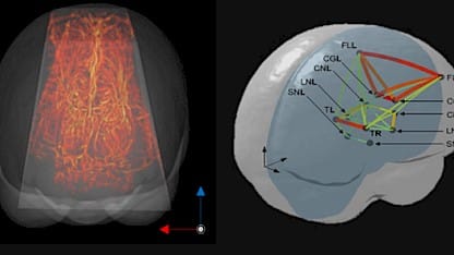
In a paper published in Nature Communications, we’ve described initial work that shows how fUS can overcome the difficulties of using existing neuroimaging tools on newborns, for early detection of neurological impairments. An interesting preliminary finding is that there may be a significant difference in functional connectivity between the brains of term and preterm babies.
Prematurity can affect the development of the brains of neonates, and result in neurological impairments of various degrees of severity, with some consequences being life-long. Examples of pathologies include autism spectrum disorders, intellectual deficiency, epilepsy, attention deficit and hyperactivity disorders. A major challenge in neonatal medicine is therefore to detect and identify the neurodevelopmental trajectory of neonates as soon as possible after birth, but monitoring their cerebral functions is difficult with existing neuroimaging tools.
Tackling the problems of brain imaging using fUS
One approach is functional magnetic resonance imaging (fMRI), and although this is the ‘gold standard’ for neuroimaging, its cost, susceptibility to motion-derived artifacts, and the cumbersome set-up make it difficult to use for longitudinal monitoring of vulnerable subjects such as preterm neonates. Two other methods – electro-encephalography (EEG) and near-infrared spectroscopy (NIRS) – can be used at the bedside, but have limited spatial resolution and imaging depth.
This is where functional ultrasound (fUS) comes in. In 2017, we demonstrated for the first time the acquisition of fUS data in human neonates. Since then, we’ve been working on methods to extract functional connectivity information from this data, because alterations in the functional connectivity can be a marker of cerebral dysfunctions.
Investigating the functional connectivity in the brains of neonates
Now, in a paper in Nature Communications, authored by a team including Iconeus staff members, we’ve described how we recorded the brain activity of neonates at rest. We then used three methods to assess the functional connectivity, each exploiting different characteristics of the data and providing complementary information.
From this, we were able to extract functional connectivity patterns, and also study how they evolved over time, and how often each pattern occurred. This is a notable advance, because although dynamic functional connectivity has been studied in adults using fMRI, very little data has been obtained on neonates so far. Interestingly, these initial results obtained using fUS in a small cohort suggest a significant difference between term and preterm babies.
The potential of fUS imaging as an imaging tool for newborns
We think that fUS imaging has a lot of promise as a tool for monitoring babies’ brain functions, because of its unrivalled sensitivity, portability and ease of use. Unlike fMRI, the examination can be performed at the bedside, simply by placing an ultrasound probe on the baby’s head.
Ludovic Lecointre, Pharm.D., CEO and co-founder of Iconeus, said:
“This study takes functional ultrasound one step further towards clinical applications, by defining advanced analysis methods to provide a detailed and robust assessment of both static and dynamic functional connectivity. We have great hopes that fUS will greatly facilitate clinical investigations, and ultimately provide a new and powerful tool in neonatal care”.
References:
J. Baranger, C. Demene, A. Frerot, F. Faure, C. Delanoë, H. Serroune, A. Houdouin, J. Mairesse, V. Biran, O. Baud and M. Tanter, Bedside functional monitoring of the dynamic brain connectivity in human neonates, Nature Communications, 2021, 12: 1080.
