New research using fUS uncovers role of minor blood vessels on the success of ischemic stroke treatment
A paper published in the journal Neuron shows that leptomeningeal collaterals are important in moderating the high blood flows that can result from treatment of ischemic stroke.
Ischemic stroke affects millions of people annually, but removing the arterial blockages that cause it does not have a high success rate. New research using Iconeus’ fUS imaging equipment, and published in the journal Neuron with Iconeus staff as co-authors, now offers an insight into why this might be the case, with minor blood vessels known as leptomeningeal collaterals shown to play a significant role in moderating blood flows following blockage removal.
The blockage of arteries within the brain, known as ischemic stroke, leads to disability and death in millions of people every year. Removal of the blockage, in a procedure known as recanalization, is the main method used to restore blood flow to the affected areas, but it often fails to work well, for reasons that are not well understood.
Now, with the help of fUS equipment from Iconeus, Professor Dr Susanne Wegener and colleagues at the University Hospital of Zürich, Switzerland, have carried out a study in mice that shows the importance of so-called ‘collateral’ blood vessels in improving the outcomes of recanalization.
Understanding the role of leptomeningeal collaterals
The study focused on leptomeningeal collaterals (LMCs), which are found on the surface of the brain, and which under normal physiological conditions have very low blood flows. But when a major vessel is blocked during an ischemic stroke, blood flow through them can increase substantially. It has previously been shown that where this takes place, it can help (at least in part) to maintain the blood flow to the areas affected by the stroke.
The new study, published in the journal Neuron, now shows that LMCs can also help to modulate the restoration of blood flows (reperfusion) following removal of the arterial blockage, helping to avoid damage to the previously blood-deprived tissues.
To achieve this insight, the authors applied a variety of analytical techniques to a mouse model in which strokes were induced by injection of thrombin. The LMCs themselves were relatively easy to study because their location near the brain surface meant that they could be imaged by conventional techniques, such as two-photon microscopy and laser speckle contrast imaging (LSCI).
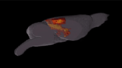
But the arterial vessels where the blockage had been induced are located deeper in the mouse brain, which is beyond the capability of these techniques. The authors therefore turned to functional ultrasound (fUS), which is able to image the entire mouse brain, and so can provide a fuller picture of blood flows, including the deeper arterial vessels.
Using fUS to study blood flows deep in the mouse brain
With the help of staff at Iconeus (who are co-authors of the article), Wegener and colleagues carried out fUS studies on anesthetized, head-fixed mice, using our Iconeus One system along with our probes and motorized platform. The resulting set of 24 stacked coronal images, each separated by 0.3 mm, enabled them to reconstruct the blood flows in the vessels within a 10 mm × 12.8 mm × 8 mm volume of the mouse brain. Further information was obtained by our microbubble-enabled ultrasound localization microscopy (ULM) technique, which provides enhanced contrast and improves resolution for even the smallest of blood vessels.
The team also took advantage of our ‘Brain Positioning System’ (BPS™) software, which by enabling automatic alignment of vascular images with the Allen Mouse Brain Atlas, helped them to enhance reproducibility.
The authors highlight the important role of the imaging technology – including our fUS instrumentation – in their research, saying: “By combining [functional] ultrasound with two-photon microscopy and LSCI, we established a unique picture of the dynamic changes in diameter and flow in both the collaterals themselves as well as the vessel segments in the [deeper arteries] that they perfuse during occlusion, as well as collateral flow, vascular integrity, and reperfusion patterns from the single-vessel level to deep brain areas.”
The importance of collaterals for preventing and treating ischemic stroke
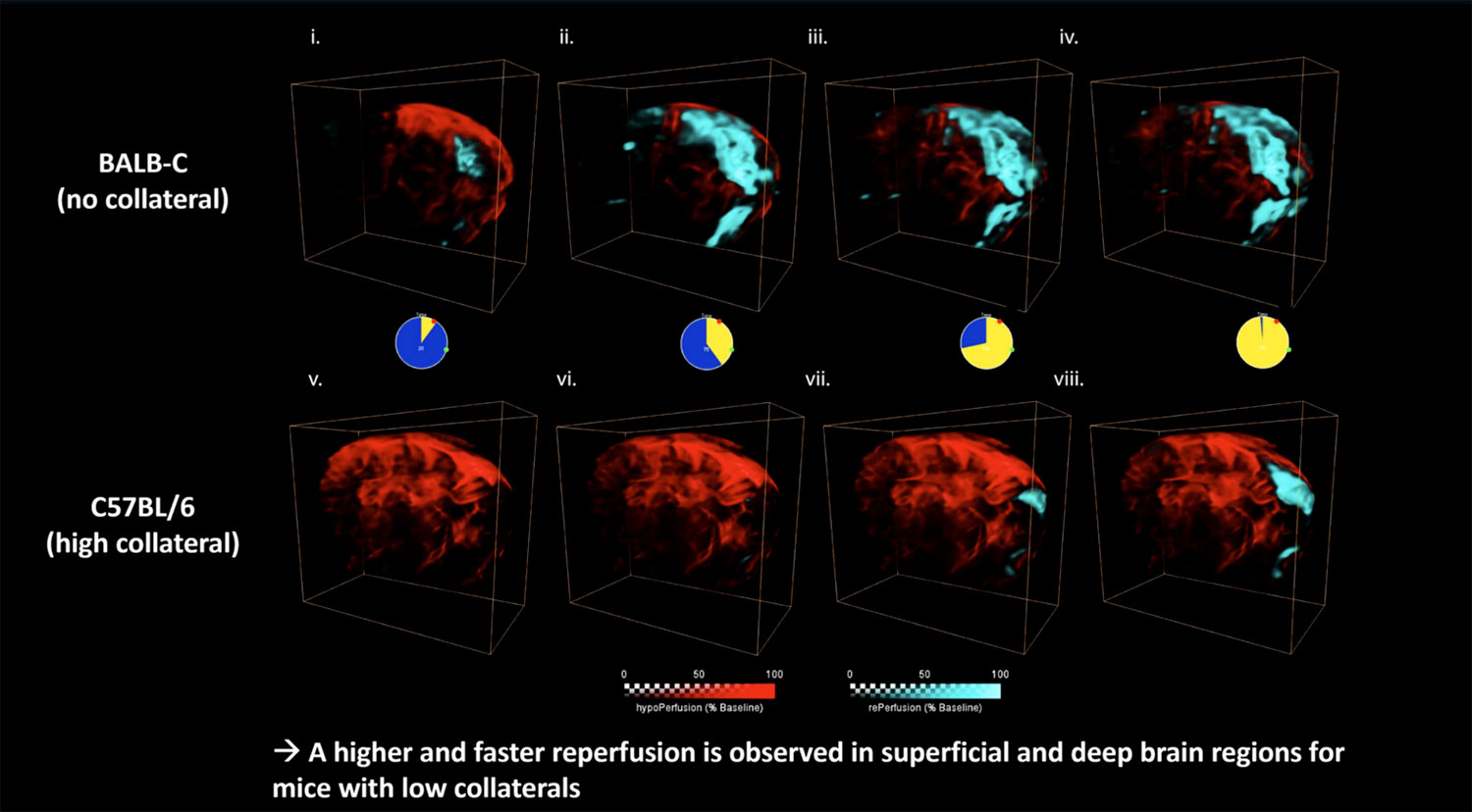
One of the key findings of this research is that reperfusion within stroke-affected areas varied greatly depending on how good the LMC network was. Significantly, mice with poor-to-moderate LMC networks showed an overshoot of blood flows in the affected region, with reperfusion taking place much more rapidly. The authors found that this faster, more extreme reperfusion resulted in a greater likelihood of more severe tissue injuries such as hemorrhaging.
When the mouse data was considered alongside clinical data also acquired as part of the study, it allowed the authors to conclude that the LMC network has an important role to play in both preventing and treating ischemic stroke. The authors say that LMC status should be considered as an indicator of vulnerable patients in stroke trials, and also that the presence of a good LMC network is “crucial” for preventing reperfusion injury arising when blockages are removed by recanalization.

Ludovic Lecointre, Pharm.D., CEO and co-founder of Iconeus, congratulates all of those involved in the research: “This is a very thorough study that sheds new light on the underlying mechanisms involved in ischemic stroke and its effective treatment by recanalization. Helping to understand the risk factors and improve treatment outcomes for ischemic stroke is something that we’re very much attuned to here at Iconeus, and we’re pleased that our functional ultrasound equipment is helping researchers tackle this important subject”.
Reference:
N. Felizitas Binder, M. El Amki, C. Gluck, W. Middleham, A.M. Reuss, A. Bertolo, et al., Leptomeningeal collaterals regulate reperfusion in ischemic stroke and rescue the brain from futile recanalization, Neuron, 2024, DOI: https://doi.org/10.1016/j.neuron.2024.01.031.