Monitoring heart and breathing rates is essential during fUS and fMRI brain imaging studies, but getting reliable measurements is fraught with practical difficulties. Now, a research team involving Iconeus has used fUS data on large-scale brain tissue motions – previously discarded during signal processing – to provide accurate information on cardiac and respiratory pulsation, both in rodents and humans, in a real-time approach termed ‘physio-fUS’.
Cardiac and respiratory rates are powerful indicators of stress, pain and disease, and it is commonplace to monitor them during any sort of in vivo study or operation on animals – for example, to check animal welfare or to ensure homogeneity of acquired data.
However, using the various methods for monitoring heart and breathing rates during fUS or fMRI neuroimaging studies on small animals is fraught with challenges. For example, electrodes remain the reference device for acquiring this data, but because they need to be implanted, they are only usually compatible with anesthetized rodents – precluding their use as part of fUS studies on awake, active animals.
‘Smart’ textiles and wireless technologies offer a solution to this, but they tend to be expensive, and the small, delicate bodies of rodents makes it difficult to position sensors so that they will provide accurate measurements for the duration of an experiment. In addition, like implanted electrodes, they further complicate an already intricate setup, and present a major challenge in synchronizing and integrating data streams from different sources.
Making use of ‘waste’ fUS data
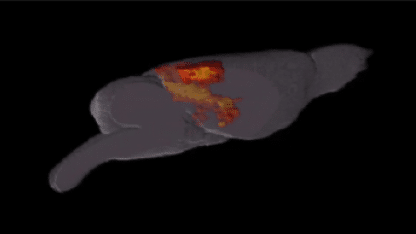
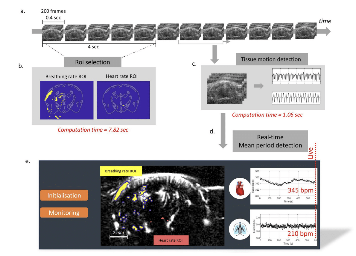
A way of overcoming these practical difficulties, termed ‘physio-fUS’, is now reported in a research article in eBiomedicine (part of The Lancet family of journals). In the article, which was published on 30 January 2025, a Paris-based team involving staff from Iconeus and Physics for Medicine Paris describe how they took advantage of the fact that breathing and heart pulsation cause periodic movements in bulk brain tissue. This is an inherent feature of all fUS studies, and in fact the ultrasound reflections it produces are typically removed during signal processing, in order to improve the quality of the blood-volume data that is the usual focus.
Realizing that this ‘waste’ data could provide useful insights, the team developed algorithms to semi-automatically identify within it separate ‘regions of interest’ in the brain that most clearly show the expected regular patterns. Having done this, simply monitoring the signal over time in these two regions then provided the desired data on the period of cardiac and respiratory pulsation.
Validation against electrode data
To validate the data acquired using this fUS-based approach, the team simultaneously took measurements using implanted electrodes, and found very good linear correlation between the two sets of values, with sub-1% errors for heart rate and breathing rate in both mice and rats.
At this point, the team had been working on previously acquired data, so they then investigated whether the method could be used while an experiment was in progress. Encouragingly, they found that the time needed to acquire a measurement is similar to the temporal resolution of fUS acquisitions. This, they say, means that it would in principle be possible to have a live read-out of heart and breathing rate, eliminating the need for separate monitoring equipment.
Assessing clinical potential
The next step was to extend this approach to humans, which the team did with the assistance of the Robert Debré Hospital in Paris. Six preterm newborns were fitted with an fUS headset, and imaging took place non-invasively through the anterior fontanelle while they slept.
The results clearly showed that information on heart rate can be extracted at the same time as fUS signals, with a 1% deviation from the ‘gold-standard’ ECG approach. However, for reasons likely related to the equipment setup, it was not possible to extract data on breathing rate on this particular occasion.
Ideas for future studies
In conclusion, the authors are optimistic about the potential of this ‘physio-fUS’ method, saying that the ability to carry out live monitoring of physiological parameters using fUS data would reduce the need for additional sensors, while also ensuring precise synchronization of data streams that up to now has been difficult to achieve.
They go on to say that on the basis of the neonate work, the method shows “strong potential” for translation to the clinic. Moreover, because of the complex relationship between physiological parameters and neuronal activity, there is a possibility that it “could help elucidate the connections between physiological rhythms and brain connectomics, neurovascular coupling, brain states, or circadian rhythms”.
Nevertheless, there is plenty of work still to be done to refine the method, including making selection of regions of interest more robust, investigating how to extract information on the breathing rate across all experimental setups, and carrying out further validation against gold-standard methods. They conclude by considering future avenues for research, which include applying the method to other commonly used modes of fUS acquisition, and assessing its compatibility with injected contrast agents, such as the microbubbles used in Iconeus’ ULM method for imaging brain vasculature.
Reference:
N. Zucker, S. Le Meur-Diebolt, F. Cybis Pereira, J. Baranger, I. Hurvitz, C. Demené, B.-F. Osmanski, N. Ialy-Radio, Valérie Biran, O. Baud, S. Pezet, T. Deffieux and M. Tanter, Physio-fUS: a tissue-motion based method for heart and breathing rate assessment in neurofunctional ultrasound imaging, eBiomedicine, 2025, 112: 105581, DOI: 10.1016/j.ebiom.2025.105581.