Iconeus One

The only fUS system designed specifically for neuroscientists

Iconeus One is an imaging system with a difference – one with the sensitivity and resolution needed to see what’s happening in the brain at the finest scales. Not only does it work on awake or moving animals, but you can even see what’s happening in real-time.

And we’re well aware that you don’t want to spend ages learning how to use it. So we’ve taken special efforts to make the software and hardware easy to use – and of course customizable to your specific needs.

What you get with Iconeus One

Here’s what’s available with our standard Iconeus One package:

Probes, robotic automation, software and more…​​

Our plane-wave functional ultrasound technology lies at the heart of the success of Iconeus One. But the performance of the whole system depends on some other innovations too, including:

The result? A combination of research-quality performance with ‘plug-and-play’ operation that makes it easy to achieve amazing results.
Picture of Dr Jeremy Ferrier

Quality products… and customer commitment​

At Iconeus, we’re committed to developing quality products that are easy to use and that stand the test of time. All our hardware and software is designed from the ground up and manufactured to the highest standards, for reliable fUS imaging – day in, day out.

Every fUS system and probe that we ship is thoroughly tested beforehand. And if there are any issues that we can’t resolve easily, we’ll just ship a new replacement machine or probe to you, so that you can carry on with your work.

“Happy to say that the Iconeus team has never failed to deliver, and always with maximum results. Thank you!”

– Artem Shatillo, MD, Charles River

Monitor brain activity in animals when they’re awake and moving

Iconeus One can be configured to provide excellent results whatever your animal is up to – whether it’s awake or asleep, freely moving or resting. This means that you’re not limited to studying anesthetized animals, as is the case with fMRI. 

And we’re familiar with devising setups for imaging of a wide range of animals. As well as rats and mice, we’ve developed probes for primates, guinea pigs, ferrets, marmosets, birds and even reptiles. You name the animal model, and we’ll work out an ideal configuration.

Head-fixed configuration in mice with a Mobile HomeCage™ from NEUROTAR™, associated with an ICONEUS transducer via a compatible headplate.

Power Doppler images obtained from an automatic scan of a rat’s brain. Voxel intensity is directly proportional to cerebral blood volume (CBV).

Take a 3D scan across a whole rodent brain in a matter of seconds

Many brain-imaging technologies are limited to a small field of view, but Iconeus One opens up new perspectives. Using our plane-wave ultrasound probes, a 2D image across a section of the brain can be obtained in about 400 milliseconds, allowing you to create videos showing changes in cerebral blood volume (CBV) even over short timescales. In addition, by using our scanning platform and XYZ+R robot, you can build up slices into a complete 3D image of a brain in as little as 20 seconds. 

In mouse studies, Iconeus One also reduces the need for pre-scanning preparation of the cranium, as the brain can be imaged directly through the skull. Other animals usually need an ‘acoustic window’ to be created, by thinning the cranium or replacing it with an acoustically transparent polymer.

Detect subtle changes in neuron activation at the sub-mm scale

The ultra-fast, plane-wave approach used by Iconeus One results in tens of thousands of ultrasound echoes being received per second. This means that multiple echoes can be combined to give a high signal-to-noise ratio, while still providing a high image framerate. The exact figures will vary depending on your application, but typically you’d be able to resolve hemodynamic changes of only 2% without averaging over different trials.

The spatial resolution typically possible with Iconeus One is as good as, or better than, the best fMRI instrument – for example, our standard 15 MHz probe generates individual volume-pixels (‘voxels’) that are 100 × 100 μm square, and 400 μm thick. That’s enough, for example, to detect signals from small vessels with blood velocities as low as 1 mm/s.

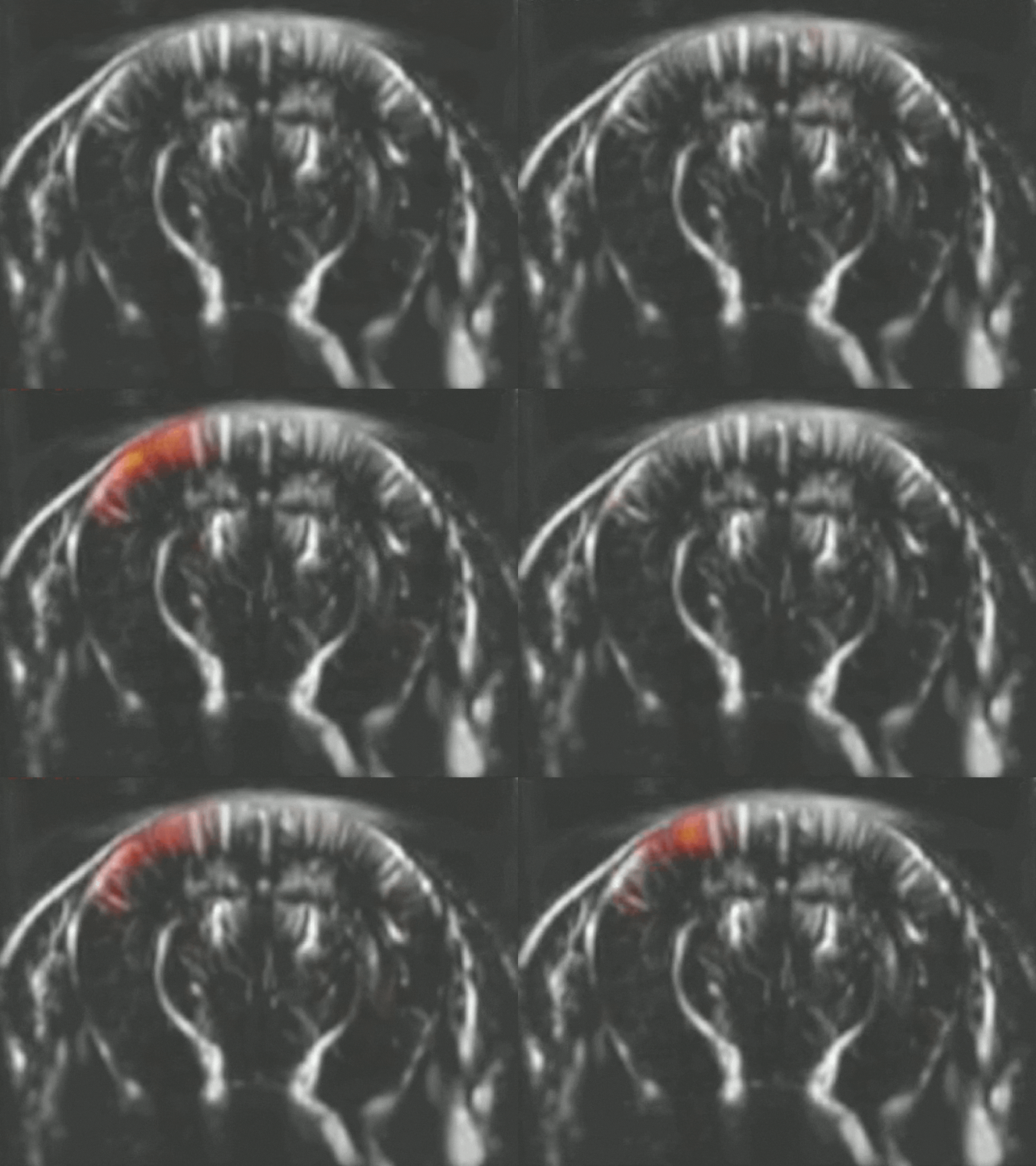
Transcranial imaging of the mouse brain during whisker stimulation.

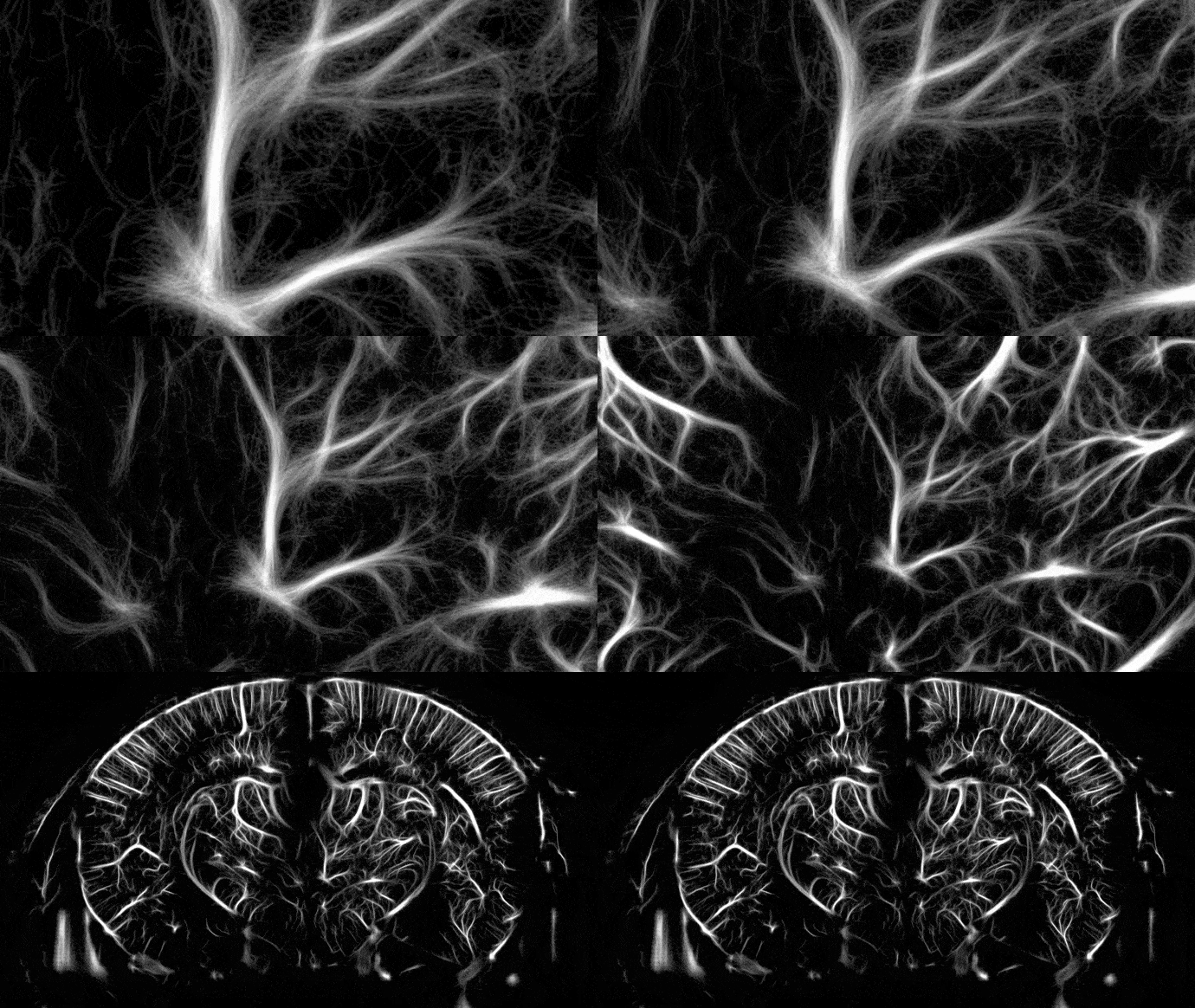
High resolution vascular map of the mouse brain (approx. –2.30 mm from Bregma) obtained with ULM. The image was reconstructed from a 5 min recording, following intravenous injection of a single bolus of microbubbles.

Generate breathtaking images of the brain microvasculature

The 100 µm spatial resolution possible with our standard Iconeus One instrument is actually just the beginning. You can now go much further with our ultrasound localization microscopy (ULM) option, which allows you to achieve resolutions as low as 5 µm, and quantify blood velocities in the mm/s range.

ULM is available as an option for Iconeus One – just ask us about it when you request a quote or book a demo.

Start seeing results immediately

Iconeus One is ready to use straight out of the box. You won’t waste your time on programming, and you don’t need to be an expert in ultrasound imaging to get impressive results. With just a few days’ training from our technical team, and with the help of software presets for rapid acquisition and processing, you’ll be generating new insights faster than you thought possible.

And because you’ll have your own system, in your own lab, there’ll be no need to book time slots on the fMRI down the corridor – your Iconeus One system will be ready for you to use, whenever and wherever you need it.

We’ve also integrated the Allen Mouse Brain Atlas into the Iconeus One software. This allows automated, accurate identification of brain regions, and opens up exciting options such as live probe positioning, standardized acquisition of large datasets, and more.

3D images of the mouse brain following whisker stimulation obtained with our inbuilt IcoStudioTM software.

fUS probes optimized for your application

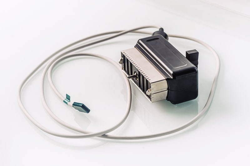
What you get from functional ultrasound depends crucially on the specifications of the probe: regular ultrasound transducers simply aren’t good enough for brain imaging. So we’ve spent a lot of time maximizing the sensitivity of our probes, minimizing their size for mobile configurations, and working out the best specifications – including optimal frequencies – for different animal models.


Ultimately, we know that as a researcher, you want to keep ahead of your field – and for that you need a functional ultrasound system that’s optimized for your animal and your application. So we’ll be delighted to hear about your work, and develop a probe and platform tailored to you.

What does Iconeus One cost?

We hope you’ll agree that functional ultrasound could offer a lot for your neuroscience research. So what does it cost?

To give you an approximate idea, our standard Iconeus One package costs about the same as a good confocal microscope… making it a fraction of the price of an fMRI scanner. And when you consider the insights that you can gain with fUS, we think that’s a pretty good deal.

Interested? Then drop us a line to find out more, have a chat about your research, and request a quote. We look forward to hearing from you.
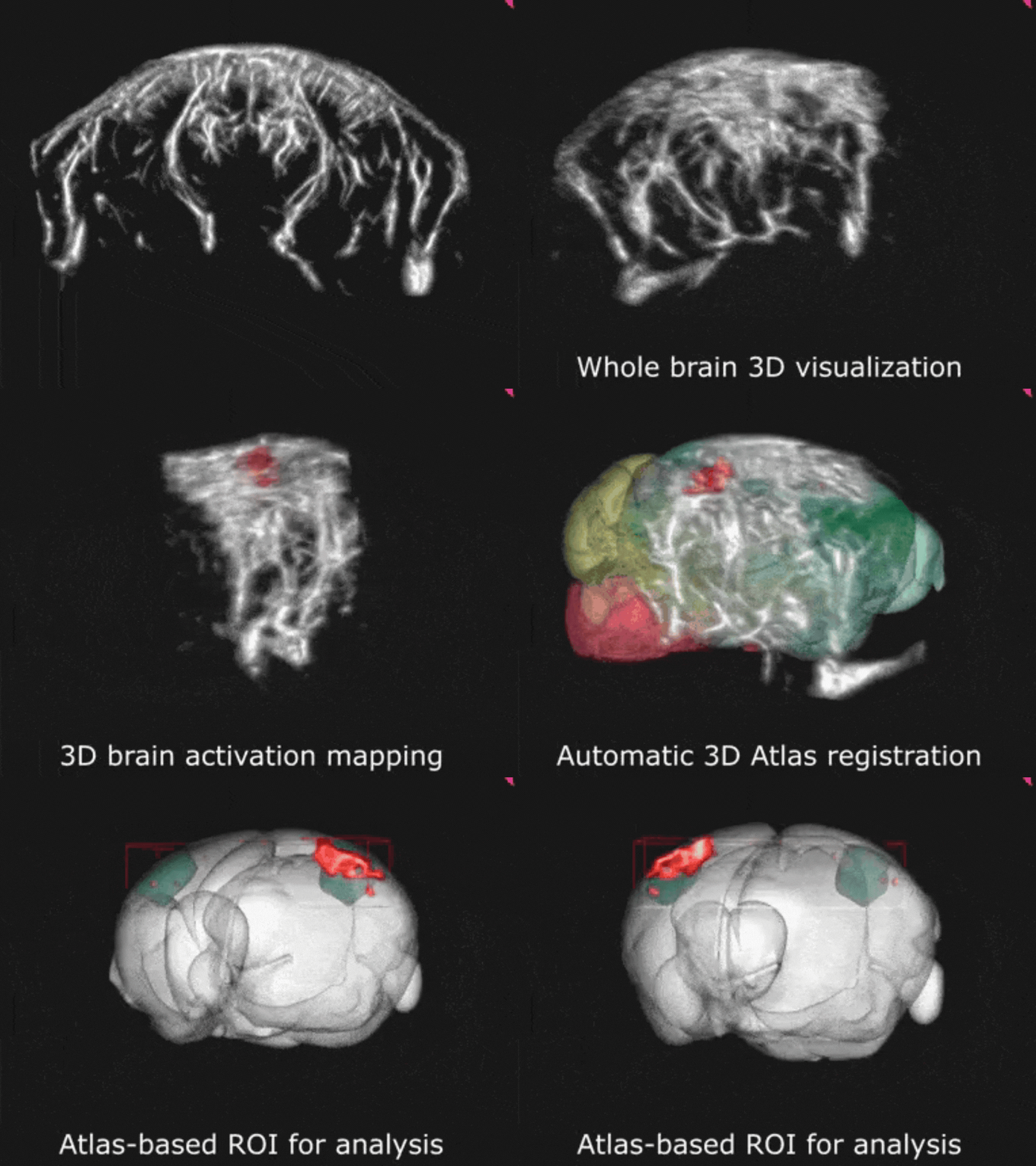
Image of participants at fUSbrain 2019
fUSbrain 2019 - Iconeus was a proud sponsor of the very first fUS brain imaging conference held in Cargèse, Corsica, with participants from prestigious institutions such as Caltech, Mayo Clinic, Charles-River and Boehringer Ingelheim.

Join the fUS community

In addition to the product itself, as a customer you’ll also benefit from being part of our network of researchers using functional ultrasound. 

Collaboration lies at the heart of our community. Through our network of Iconeus One users, we share tips, advice and ideas, while workshops bring together researchers to advance neuroscience and discover new ways of using functional ultrasound.

Get in touch