
What is functional ultrasound (fUS)?

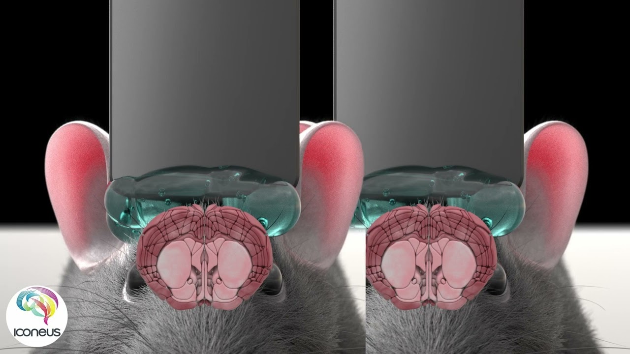
How does functional ultrasound work?
What is fUS?
How does it work?
A window on brain activity
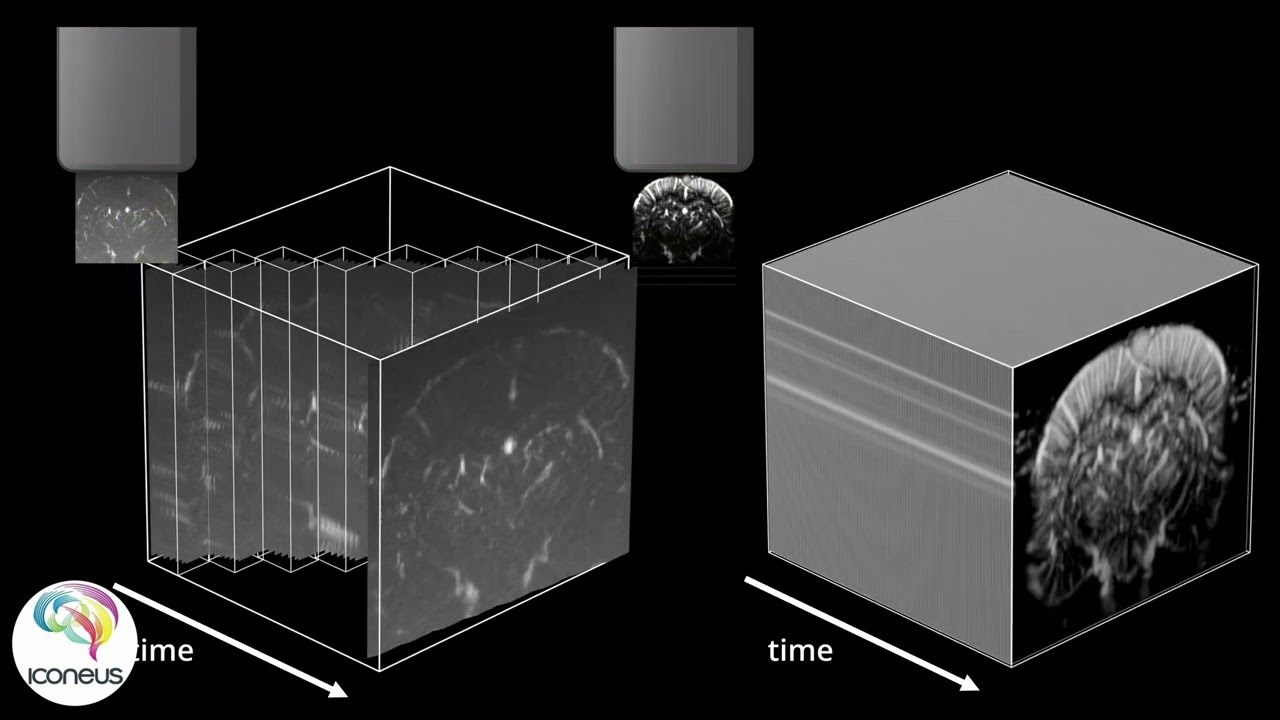
The concept of functional ultrasound was presented in a high-profile paper co-authored by our founder, Mickael Tanter (Macé et al., Nature Methods, 2011). Because fUS uses a succession of ultrasound ‘plane waves’, rather than a beam focused on a point, echoes are received from an entire 2D plane in one go. This improves on conventional ultrasound imaging by avoiding the need to acquire data from small volumes sequentially.
But the success of fUS also depends crucially on the rate at which those plane-wave pulses can be generated. Because we employ ultrasound technology capable of generating pulses at about 20 kHz, many responses can be combined into one image. This improves the signal-to-noise ratio and so vastly increases sensitivity.
Every ultrasound pulse transmitted gives rise to an echo (backscatter) from the bodily tissues. After subtracting unwanted low-frequency ‘quasi-static’ echoes, we are left with the higher-frequency signal due to the moving red blood cells alone. The mean intensity of this signal is known as the ‘Power Doppler’ value, which is proportional to the number of red blood cells in a unit volume, and hence to the cerebral blood volume (CBV).
The fact that brain activity is closely synchronized with the dilation of the blood vessels supplying the neurons involved (the neurovascular coupling) means that the CBV measured by fUS can be used as a surrogate for neuronal activity.
Therefore, with a bit of behind-the-scenes number-crunching, the complex datasets generated from an fUS scan can be converted into easy-to-understand images (and videos) showing patterns of brain activation – as they happen.
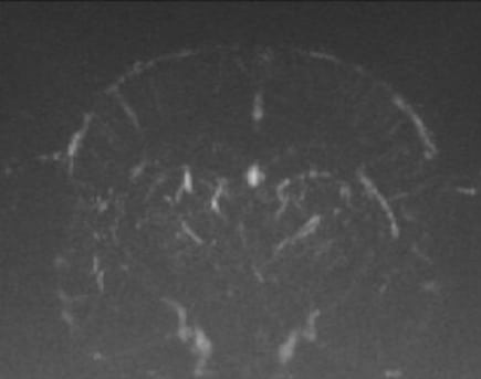
Conventional ultrasound vs. functional Ultrasound (fUS)
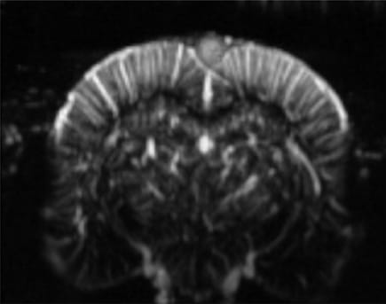
Functional ultrasound represents a major leap forward in neuroimaging, offering enhanced sensitivity and precision compared to conventional ultrasound. Use the slider on the right to compare images acquired with conventional ultrasound (left) and fUS (right).
Interested in learning more about the difference between conventional ultrasound and fUS? Read our perspective article
Key differences and advantages of fUS:
- 100-Fold increase in sensitivity: fUS boasts a 100-fold increase in sensitivity compared to conventional ultrasound, enabling it to detect minute physiological changes that were previously undetectable.
- Captures slow blood flows: Unlike conventional ultrasound, fUS can capture blood vessels with velocities as low as 1mm/s. This includes arterioles and venules, providing detailed insights into microvascular flow.
- Direct rCBV measurement: The signal in fUS is directly proportional to relative cerebral blood volume (rCBV). This direct correlation allows for more accurate mapping of blood flow and volume changes within the brain.
- Single-trial detection of hemodynamic changes: The high sensitivity of fUS makes it possible to detect hemodynamic changes in a single trial. This capability is crucial for real-time monitoring and analysis of brain activity, including brain-machine interface.
- High Signal-to-Noise ratio: Plane-wave imaging allows for advanced clutter filtering methods, yielding exceptional SNR. This makes fUS a powerful tool for studying subtle physiological changes with great accuracy.

Discover Iconeus One

Reproduced from Boido et al., Nature Communications, 2019 (licensed under CC BY 4.0).
Better than BOLD-based fMRI
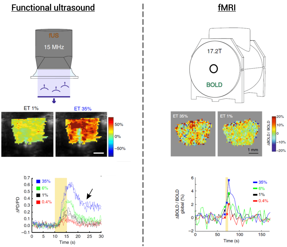
Functional magnetic resonance imaging (fMRI) using the blood-oxygen-level-dependent (BOLD) response is the ‘gold standard’ for deep brain imaging in humans. But its restricted spatial and temporal resolution makes it of limited use when looking at small animals in preclinical settings (especially mice). Also, the small responses generated by BOLD mean that you need to take an average of many experiments to get an acceptable signal-to-noise ratio.
Functional ultrasound using Iconeus One provides an alternative approach, by offering:
- High spatial resolution (down to 100 µm)
- High temporal resolution (as low as 100 ms)
- High-sensitivity (detect CBV changes as low as 2% compared to baseline, thanks to its high signal-to-noise ratio)
The result is performance that’s as good as, or better than, the best fMRI systems – even minor blood vessels in small mammalian brains can be imaged with a single scan. Not only that, but the size of the probes allows animals to be imaged while they’re awake and moving.
Complementary to other techniques
In contrast to fMRI and the strong magnetic fields it requires, fUS has little (or zero) impact on tissues or equipment. It’s therefore straightforward to combine with other modalities such as electro-encephalography (EEG), multi-electrode arrays, positron emission tomography (PET), optical imaging, or optogenetics.
This means you can easily obtain a detailed understanding of brain vasculature and blood flows at the same time as carrying out electrophysiology, metabolism, optogenetics, guided injections and more… and so gain better research insights.