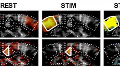
Functional ultrasound is a rapidly-evolving field of neuroscience research, and evidence of this is provided by three articles, authored by teams including Iconeus founders, which report major advances in both the fundamental aspect of the fUS method and its applications for preclinical research.
Since its introduction in 2011, functional ultrasound (fUS) imaging has rapidly progressed as a field of study. This is demonstrated by the publication, just a few days apart, of three papers in the high-impact journals PNAS and Nature Communications, which describe the methodology and applications of the fUS.
Elucidating the origin of fUS signals measured during brain activation
The first study[1] is authored by a team led by Serge Charpak at the Institut de la Vision (Sorbonne Université), and including Iconeus founder Mickael Tanter. The research investigates the link between the ultrasound signal recorded in a single voxel and the neuronal activity within that same voxel, by co-registering fUS and bi-photon microscopy measurements. This allowed the team to establish transfer functions that enable prediction of fUS signals from sensory stimulation.
Discovering a ‘default mode network’ in the mouse brain
In the second publication,[2] a team led by our co-founder Zsolt Lenkei (affiliated to the Institute of Psychiatry and Neuroscience of Paris, and the Physics for Medicine Paris Laboratory) have observed for the first time in mice the functioning of a neural network that is known in humans as the ‘default mode network’, and which is affected by brain disorders such as Alzheimer’s disease. Studying this network in mice could therefore enable the development of new therapeutic drugs for neuropsychiatric disorders using a translational approach.
Precisely mapping the visual cortex in primates
The third study[3] is a collaboration between the teams of Pierre Pouget at the Paris Brain Institute and Serge Picaud at the Institut de la Vision (Sorbonne Université), with assistance from Iconeus founders Thomas Deffieux and Mickael Tanter. In this work, the authors create activation maps of the primary visual cortex in primates at unprecedented precision, enabling the differential activation of layers of the primary visual cortex across its entire depth to be distinguished.
References:
[1] A.-K. Aydin, W.D. Haselden, Y. Goulam Houssen, C. Pouzat, R.L. Rungta, C. Demené, M. Tanter, P.J. Drew, S. Charpak and D. Boido, Transfer functions linking neural calcium to single voxel functional ultrasound signal, Nature Communications, 2020, 11: 2954.
[2] J. Ferrier, E. Tiran, T. Deffieux, M. Tanter and Z. Lenkei, Functional imaging evidence for task-induced deactivation and disconnection of a major default mode network hub in the mouse brain, Proceedings of the National Academy of Sciences, U.S.A., 2020, 117: 15270–15280.
[3] K. Blaize, M. Gesnik, F. Arcizet, H. Ahnine, U. Ferrari, T. Deffieux, P. Pouget, F. Chavane, M. Fink, J.A. Sahel, M. Tanter and S. Picaud, Functional ultrasound imaging of deep visual cortex in awake non-human primates, Proceedings of the National Academy of Sciences, U.S.A., 2020, 117: 14453–14463.
