
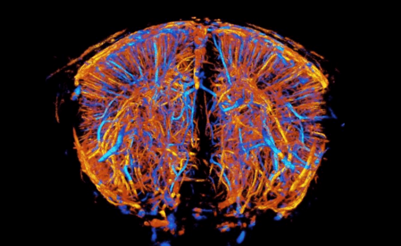
Using matrix probes for real-time 3D Ultrasound Localization Microscopy (ULM) brain angiography limits the field of view. But turning to Iconeus’ row-column addressed (RCA) probes allows super-resolution transcranial imaging of the whole mouse brain, for automated in-depth vascular classification.
The function of brain cells is intimately linked to the transport of oxygen and nutrients through the vascular network – meaning that being able to image this network has the potential to improve our understanding of a range of brain pathologies. For example, minor changes in vessel structure and function could be used to monitor the onset and development of conditions including small-vessel diseases, neurodegenerative disorders, vascular stroke, and brain tumors.
However, our understanding of the brain microvasculature is hampered by the difficulty in studying it in the necessary detail. In preclinical animal models, even distinguishing between arteries and veins is not straightforward using current techniques. The main in vivo modalities, magnetic resonance imaging (MRI) and computed tomography (CT), have limited resolution and are not easily applicable to whole-brain scanning. And although some ex vivo visualization techniques – such as phase-contrast tomography and transparency-inducing ‘optical clearing’ protocols – can overcome these difficulties, they are inherently incapable of providing the information on blood flows that is useful for vessel classification, or allowing longitudinal studies.
ULM: From two to three dimensions
Now, a team based at Iconeus and Physics for Medicine Paris has shown how an existing in vivo ultrasound-based technique for microvascular imaging can be extended from two dimensions to three dimensions, delivering high-resolution information on vessel structure and blood flows, without compromising field of view or the ease of lab implementation.
The work, reported in the Journal of Cerebral Blood Flow and Metabolism, involves Ultrasound Localization Microscopy (ULM). This technique was first reported by Iconeus’ founders in 2015, and uses functional ultrasound (fUS) imaging to track individual microbubbles as they move throughout the brain microvasculature, with the contrast enhancement from the microbubbles enabling transcranial imaging in rodent models.
Since then, a further study has shown how the imaging capability of ULM can be extended from two dimensions to three dimensions using 2D transducer arrays (so-called ‘matrix probes’). However, there are technical, data-handling, and cost challenges in scaling-up these probes to capture 3D images on a whole-brain scale, limiting access to the technology.
Dynamic switching of the transducers using multiplexers is one solution, but this reduces the effective volume rate, compromising microbubble tracking efficiency and overall image quality.Instead, in the current work, the Paris team have turned to the latest generation of row-column addressed (RCA) probes from Iconeus. Like other RCA probes, these combine all the inputs/responses for elements in a given row or column, which reduces the channel count and so overcomes the scaling constraints of matrix probes, in which every element is separately ‘addressable’. But, uniquely to Iconeus, they use patented XDoppler technology to cross-correlate the orthogonal datasets and so reduce the ‘sidelobe’ image artefacts that limit the usefulness of RCA technologies.
Super-resolution, brain-wide imaging
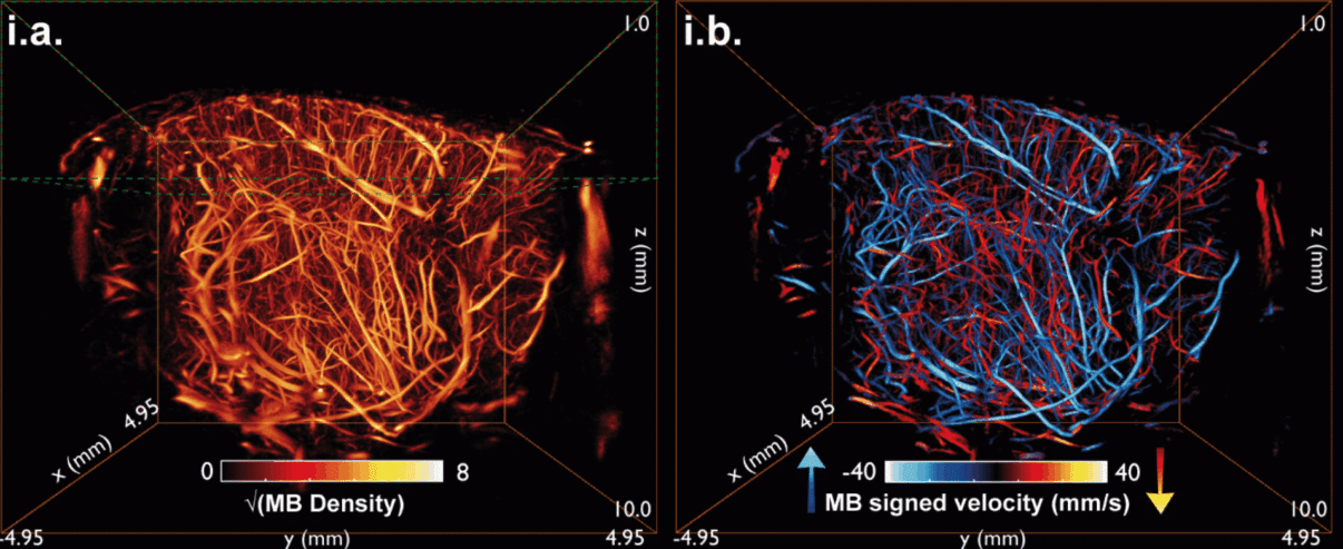
In the paper, the researchers, led by Iconeus’ Scientific Advisors Dr Mathieu Pernot and Dr Thomas Deffieux, demonstrate how these next-generation RCA probes can be coupled with ULM to image the mouse brain microvasculature in three dimensions, and how the resulting high-sensitivity data can be processed to classify blood vessels.
The team injected five mice with SonoVue microbubble contrast enhancer, and imaged them using an IcoBright-4D RCA probe connected to a 256-channel Iconeus One Xcel instrument, positioned with the aid of system’s four-axis motor module and its real-time power Doppler imaging mode. The entire ULM acquisition sequence, numbering 200,000 volumetric images in 400 blocks of 0.5 s each, was acquired over a period of 10 minutes.
Spotting patterns in microvascular data
With data acquisition complete, the researchers then developed an automated framework to classify the imaged blood vessels based on both flow dynamics and vascular topology.
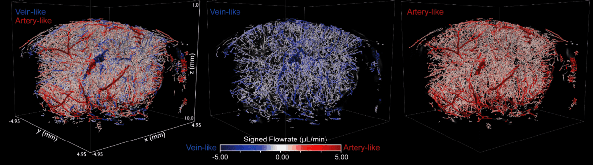
First, blood flow direction and velocity information derived from microbubble tracking were used to orient the vascular network. Changes in flow rate along vascular segments provided additional cues: a reduction signified artery-like behavior, while an increase signified vein-like behavior. In parallel, the vascular network was represented as a graph, where branching patterns were analyzed: diverging structures were associated with artery-like behavior, while converging structures were associated with vein-like behavior.
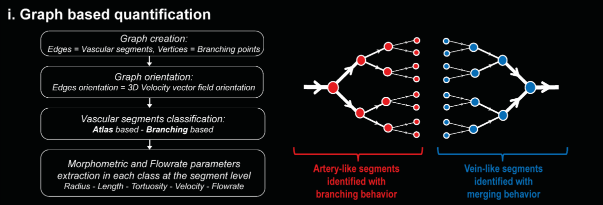
Encouragingly, the two approaches showed 85% agreement in a cortical region with a relatively simple vascular structure. This, say the authors, supports the use of the graph-based branching categorization algorithm to classify blood vessels even in regions where the vasculature is highly complex and where the easily-extractable axial blood velocity (parallel to the line of ultrasound transmission) is not a reliable indicator of vessel type.
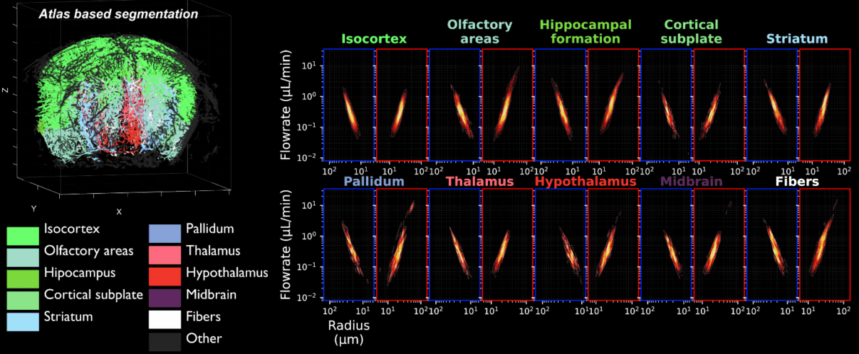
In addition, by combining this classification with an atlas brain segmentation, authors managed to extract flow rate and morphometric metrics in different brain regions automatically, for both artery-like and vein-like vessels.
Deep characterization of the whole mouse brain
The authors say that, by making true volumetric ULM more achievable in practice, this work marks a substantial advance in neurovascular imaging. Dr Adrien Bertolo, a member of the Iconeus team and first author on this study, said: “Up to now, expanding the super-resolution capabilities of ULM to three dimensions has been limited by the capabilities of matrix probes, which although offering amazing performance, can be computationally intensive and are difficult to scale up to larger volumes”.
“But by using our row-column addressed probes alongside ULM, it’s possible to acquire high-resolution, high-sensitivity vascular data across the whole mouse brain while keeping the number of channels below 256, and without using additional electronics. And that’s at the same time as maintaining the volume rates needed to acquire data with high spatiotemporal resolution – data that, as we’ve shown here, can be used to characterize blood vessels in the brain in greater depth”.
Dr Jeremy Ferrier, Head of Product Development at Iconeus, says that this new ‘RC-ULM’ approach has numerous promising applications: “There are exciting possibilities for researchers across the preclinical arena – including investigating high-throughput microvascular phenotyping in genetic mouse models, studying how microvascular networks reorganize across disease progression, and uncovering the relationship between microvascular structure, blood flow and brain function”.
“In addition, looking towards future clinical applications, one could envisage using this approach to investigate the microvasculature in surgical procedures that require removal of a section of skull, such as in tumor resection”.
Dr Bertolo concludes by considering some of the areas for future work. “As the first study that’s combined our RCA probes with ULM, there are lots of avenues to pursue. We’d like to investigate vessel pulsatility in the data, improve clutter filtering of slow-moving microbubbles to allow imaging of capillaries, and also use deep learning and online GPU-based tracking algorithms to further reduce computational intensity. It’s going to be an exciting time for brain imaging!”.
Reference:
A. Bertolo, J. Ferrier, O. Demeulenaere, A. Dizeux, T. Delaporte, B. Osmanski, M. Tanter, M. Pernot and T. Deffieux, In vivo microvascular flow quantification in the mouse brain using Row-Column Ultrasound Localization Microscopy and directed graph analysis, Journal of Cerebral Blood Flow and Metabolism, 2026,
DOI: 10.1177/0271678X261438569