Laser speckle contrast imaging (LSCI) has long been the gold-standard method for assessing functional hyperemia in preclinical models, but it suffers from limited imaging depth. A team at Oklahoma University has now explored functional ultrasound (fUS) as an alternative imaging approach in the mouse, finding a strong correlation between the two methods, and highlighting the value of the improved depth of field achievable with fUS.
Cerebral functional hyperemia – the stimulus-evoked increase in brain blood flow – is a key component of the neurovascular coupling. It’s therefore vital for healthy brain functioning, making it a very useful marker for various types of vascular cognitive impairment, including vascular dementia and Alzheimer’s.
Functional hyperemia can be imaged in various ways, of which laser speckle contrast imaging (LSCI) is the most widely used. LSCI works by detecting the characteristic ‘blurring’ effect that results when coherent light is reflected from moving red blood cells, and benefits from a simple setup and high resolution. However, a major drawback is that it cannot reliably penetrate deep tissues.
Researchers at University of Oklahoma Health Sciences Center in Oklahoma City, USA, have now shown that functional ultrasound (fUS), by allowing a greater depth of field, is an appealing alternative to LSCI for assessing functional hyperemia in a mouse model of aging.
Comparing LSCI and fUS imaging
Writing in the journal NeuroImage, a team led by Professor Stefano Tarantini and Professor Shannon Conley first implanted a polymeric cranial window into young (3–6 months) and aged mice (20–22 months). Each cohort was then divided into two groups – one that was imaged using LSCI and fUS straight away, and the other that was imaged after 14 days. The latter cohort was found to give more reliable responses, and so it was this group that was used to draw the main conclusions from the rest of the study.
In both LSCI and fUS experiments, baseline imaging of the contralateral barrel cortex was followed by 30 s of whisker stimulation on alternating sides of the face, and a further acquisition in order to determine the evoked functional hyperemia.
fUS imaging used an Iconeus One system, coupled to an IcoPrime-4D MultiArray probe, which is optimized for high-sensitivity 3D brain-wide imaging in the mouse. To ensure that the fUS results aligned with those acquired using LSCI, the researchers used the ability of Iconeus One to automatically and precisely map acquired data onto the Allen Mouse Brain Atlas.
Strong correlations
The main finding, say the authors, was “a strong positive correlation between LSCI and fUS” (with a Pearson r correlation coefficient of +0.92). They also compared functional hyperemia in young and aged mice, and noted, as expected, a significant decrease in values compared to the older cohort, from about 10% to 4% using LSCI, and from about 15% to 4% using fUS.
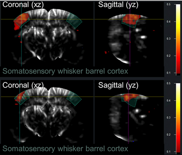
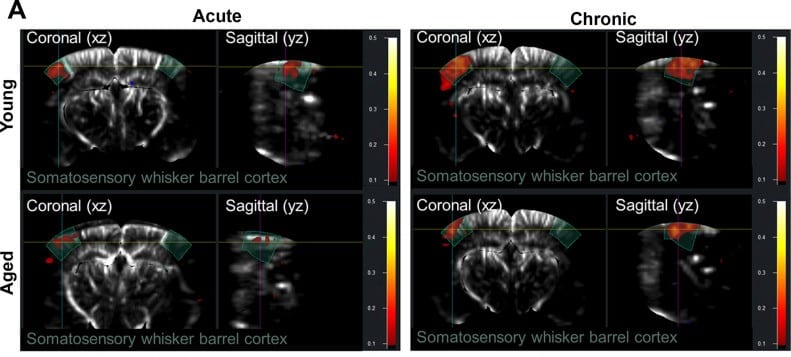
Frontal and sagittal views of the mouse brain acquired using fUS, showing a comparison of functional hyperemia responses to whisker stimulation, 14 days after the animals were fitted with a cranial window. The decrease in response between (top) young and (bottom) aged animals is apparent.
The researchers suggest that the observation that the values for young mice are substantially higher with fUS indicates that “fUS may be a more sensitive measure of [functional hyperemia] than LSCI”. They also suggest that this sensitivity, combined with a better ability to monitor at depth, “likely contribute to the superior performance of fUS in detecting dynamic hemodynamic changes compared to surface-focused techniques like LSCI”.
In a supplementary part of the study, the authors used the microbubble-enabled ultrasound localization microscopy (ULM) technique with Iconeus One to enhance the imaging contrast still further and enable high-resolution mapping of the vascular density. They also carried out a comparison against data acquired using post-mortem immunofluorescence imaging of the vasculature, again finding a strong positive correlation (Pearson r = +0.82).
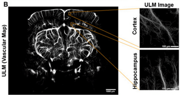
Representative ULM vascular map (left), and representative 500 µm2 sampled regions used for comparison with LSCI (right).
fUS: A transformative tool for studying cerebrovascular health
A clear outcome of the study is that fUS provides results closely matching those obtained using LSCI. This, say the researchers, positions the technique as a “reliable alternative” to LSCI for preclinical research, enhancing the prospects for its successful translation to the clinic.
They also point out that the two techniques have a degree of complementarity, saying that “fUS offers greater imaging depth and resolution, making it particularly advantageous for detecting changes in smaller vessels”. Another advantage of fUS is that, once the polymeric window is in place, the excised skin can be restored without loss of image quality, making longitudinal studies much easier.
They conclude with a positive assessment of the value of fUS for studying functional hyperemia specifically and cerebrovascular conditions in general, saying that fUS is positioned as a “transformative tool for studying cerebrovascular health, particularly in age-related dysfunctions such as cognitive decline and dementia”.
Reference:
J. Pinckard, S. Negri, C.A. Huston, M.A. Bickel, M.L. Vance, M. Milan, C.L. Hibbs, M. Budda, S.S. Chandragiri, K. Pipkin, S. Tarantini and S.M. Conley, Functional ultrasound as a quantitative approach for measuring functional hyperemia in aging models, NeuroImage, 2025, 316: article no. 121313, https://doi.org/10.1016/j.neuroimage.2025.121313.