Vascular imaging
The sensitivity of fUS to blood flow means that you can visualize the whole vascular tree, from major cerebral arteries down to minor arterioles, quickly and easily.
Such detailed blood vessel imaging has applications in a range of situations – including assessing changes in vasculature that take place during Alzheimer’s disease and aneurysm, monitoring ischemia and reperfusion in stroke, studying cerebral flow autoregulation, and investigating the response of brain tumor vasculature to therapeutic treatment.

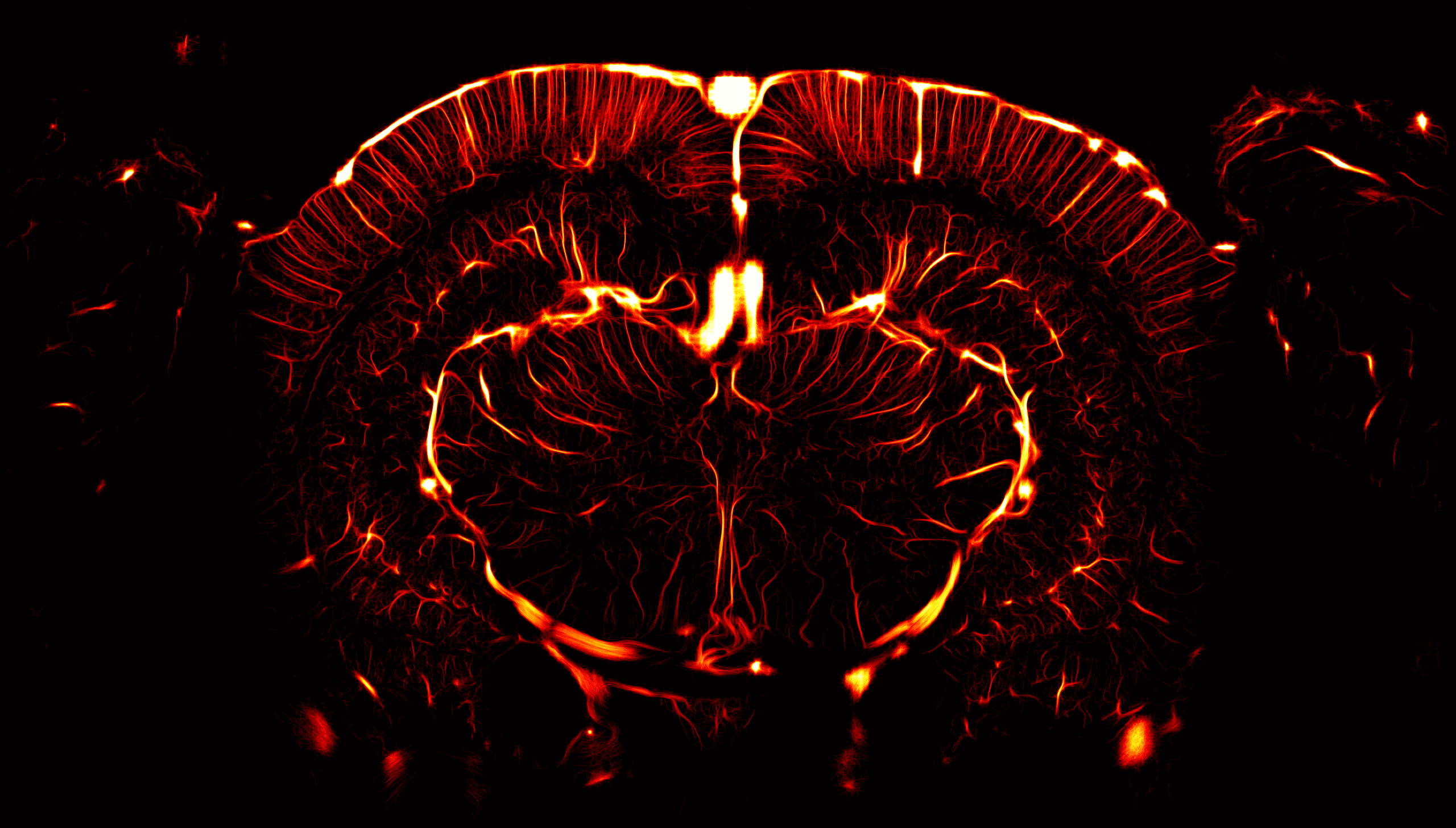
Carrying our ultrasensitive angiography
Monitoring changes in blood volumes during stroke
The combination of functional ultrasound with tomographic reconstruction offers the prospect of fine-grained 3D imaging of blood vessels in the brain, within a remarkably short time-frame. This method – first demonstrated in the rat (Demené et al., NeuroImage, 2016) – provides an isotropic spatial resolution as low as 100 μm, and allows vessels with blood velocities down to 1 mm/s to be imaged.
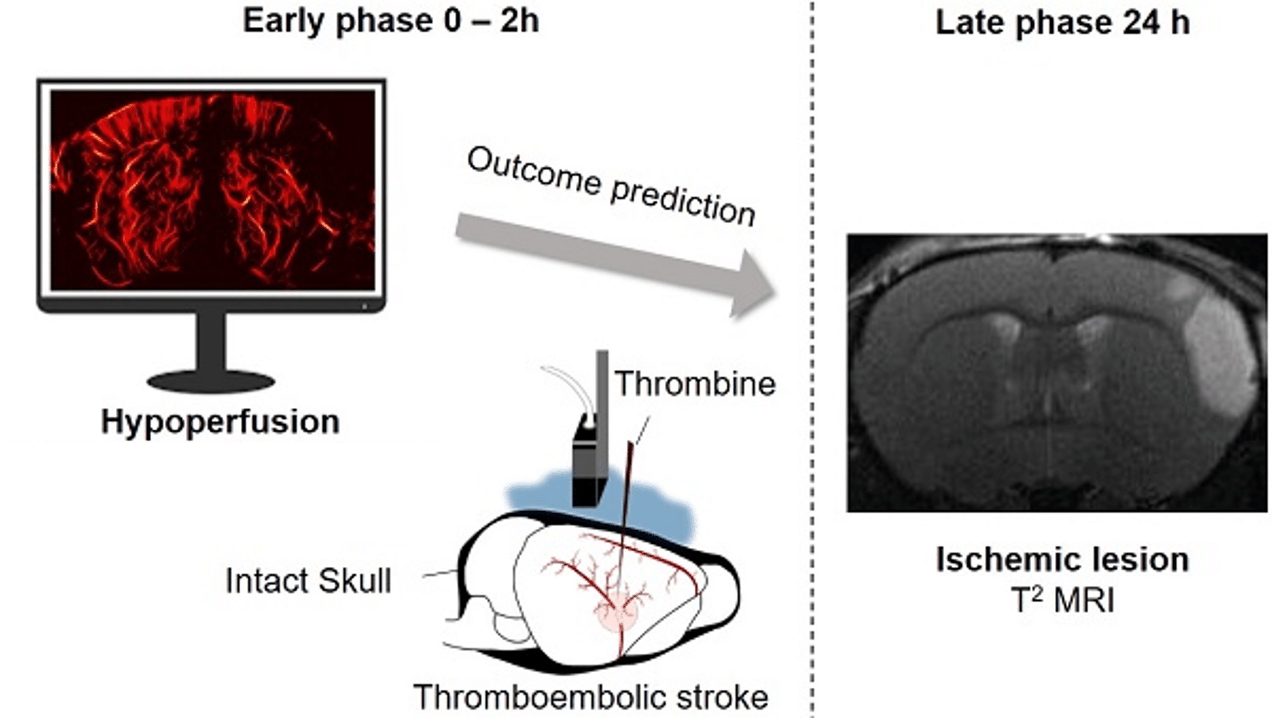
The high spatio-temporal resolution of fUS makes it valuable for monitoring changes in blood vessels in stroke models. A good example of this is an early study in the rat (Brunner et al., Journal of Cerebral Blood Flow & Metabolism, 2018), where fUS was used to characterize regional differences in blood volume in an ischemic area in the rat, during transient occlusion and subsequent reperfusion.
The later study shown in the figure provides a proof-of-concept for the additional resolution possible by using fUS with Ultrasound Localization Microscopy. In this collaboration between PhysMed and the team of Professor Denis Vivien at Normandy University, it was found that imaging of cerebral perfusion correlates with ischemic stroke outcomes and responses to treatment in mice.
Mapping blood vessels at ultra-high resolution
The concept of ultrasound localization microscopy (ULM) was published by our founders in 2015 (Errico et al., Nature, 2015), and allows amazingly detailed images of the brain microvasculature to be acquired.
In a similar way to PALM and STORM in optical microscopy, ULM uses ultrafast imaging to track injected gas microbubbles, and to reconstruct images of the brain vasculature down to the capillary level. What can be achieved is impressive – we can quantify blood velocities in the mm/s range, and can improve the resolution from 100 µm to about 5 µm. You can even track individual microbubbles as they travel through the brain’s blood vessels. ULM is available as an option on Iconeus One – please ask us about it when you call us to discuss your application.

Iconeus One
The only fUS system designed specifically for neuroscientists
Iconeus One is an imaging system with a difference – one with the sensitivity and resolution needed to see what’s happening in the brain at the finest scales. Not only does it work on awake or moving animals, but you can even see what’s happening in real-time.
Contact us
Interested by what you’ve read about functional activation mapping using fUS? Talk to one of our specialists about your application.
Other applications